強化毛囊、提升存活率

是什麼「殺死」了移植的毛囊?

移植毛囊從供區到脫髮區,不代表一定會成功生長。一旦毛囊受損,就無法再次生長。因此,每一步手術細節都需精準無誤,才能提升成活率。平均 85%–92% 毛囊在植入後能成功存活並長出新髮。
專業提醒:毛囊移植是否成功,並非「種下去就會長」那麼簡單。
每一步操作 —— 從提取、保存、到植入與術後管理,皆需精準控制,才能提升毛囊成活率與自然外觀。

造成毛囊死亡的因素主要分為兩類:X 型損害因子(X-Killing Factors); H 型損害因子(H-Killing Factors)。

X 型毛囊損害因子(X Graft Killing Factors)

“X” 代表「未知因素」,此概念最早由澳洲的 Dr. Shield 於 1984 年提出,用以解釋某些毛囊移植後無法生長的情況。初期僅佔失敗案例的 0.5–1%;隨植髮技術標準化後,大多數不理想成果其實都與 X 因素有關。我們認為這些「未知因素」其實是手術過程中及術後造成的生物性損害。

常見 X 因素包括:

我們的觀點:這些「未知因素」其實是手術當中或術後造成的生物性損傷,可透過經驗、技術與術後管理加以預防。

  • 未能辨識或錯過的 H 因素
  • 再灌流缺血損傷(Reperfusion Ischaemic Injury)
  • 毛囊自體排斥(Auto-rejection of Grafts)
  • 毛囊脫離體外時間過長,導致細胞缺氧與脫水;
  • 缺乏術後再生支援,例如 ATP 噴劑或低能量激光療法。

H 代表「Human(人為因素)」- H-Factor 是指由操作人員疏忽、病人習慣或術中技術問題所導致的毛囊損傷
可在手術前、手術中、或術後發生。因此,植髮結果無法 100% 保證,惟透過專業團隊、正確流程與完善管理,可大幅降低風險。

術前 H 因素:潛在體質或生活習慣風險

可能影響毛囊存活的身體狀況與生活習慣包括:

  • 頭皮血液循環不良(供應區或種植區);
  • 受傷或疤痕皮膚需移植(如燒燙傷、外傷);
  • 吸煙習慣(尼古丁影響血流與癒合);
  • 糖尿病未控制良好(影響傷口癒合與微循環)。
術中 H 因素:毛囊在手術中的潛在損傷

即使手術順利,若技術不當,也可能損害毛囊:

  • 毛囊脫水( 長時間暴露在空氣中 );
  • 過度去除毛囊周邊支持組織( 保護力下降 );
  • 冷凍傷害(保存液或操作溫度不當);
  • 毛囊斷裂(Transection)- 毛囊被切斷而無法成活;
  • 化學性損傷(毛囊保存期間);
  • 缺血再灌流損傷。
術後 H 因素:

移植後的毛囊受到損傷;

  • 未依照指示行事;
  • 未能建立新的微循環;
  • 移植毛囊受到外力創傷;
  • 感染。

毛囊是有生命的組織 – 當毛囊被移植到新位置後,體內會發生一連串生物化學變化,理解這些自然組織與化學過程,有助於提升毛囊存活率與最終效果。

植髮株從供區取出後,會被暫時放置於保存液中,等待植入受區。在此期間,原有的血液供應已被切斷,氧氣迅速耗盡,廢物也開始累積。
因此,植髮株能否成功從受區獲取養分並重新建立血管循環(再血管化),將直接決定其存活率。

傷口癒合階段的生理反應,一旦植入完成,身體會啟動自我修復機制,出現一連串細胞反應與生理變化,包括:
– 水腫(Edema);
– 泛紅(Erythema);
– 原有頭髮脫落(Shedding);
– 新生頭髮長出(Regrowth);
– 毛囊再血管化(Revascularization)。

組織學變化

  • 第 1 天:出現輕度腫脹(水腫)。
  • 第 3 天:大量嗜中性白血球湧入,開始清除異物。
  • 第 7 天:巨噬細胞參與修復與清除壞死組織。
  • 第 14 天:膠原蛋白與纖維母細胞活躍,組織進入癒合期。
  • 第 21-28 天:炎症逐漸減退,新血管生成完成,毛囊穩定存活。
  • 膠原蛋白 Collagen III 與 IV:從手術第一天持續表現至第六個月

本頁內容連結

毛囊存活率決定植髮成敗

在美絲植髮,我們始終堅持一個核心原則:「數量不是重點,品質才是關鍵。」每一個步驟皆以 提升毛囊存活率 為最高目標,確保每一根移植的毛囊能健康生長、持久自然。

毛囊存活率的醫學基礎與臨床意義 : 人體出生後,毛囊數量固定,無法再生;後枕部的永久毛囊(供區)為不可替代資源,許多人期待「毛囊複製技術」能解決資源限制,但至今仍未實現。因此,植髮時減少毛囊損傷與浪費, 是目前最重要的課題。

超過 50 年的臨床研究結論 – 過去半個世紀以來,全球已有眾多研究深入探討植髮後毛囊的實際存活率與生長百分比,主要結論如下:

  • 毛髮是可移植的器官(Hair is an organ)。
  • 像器官一樣,毛囊離開人體後必須妥善保存,否則會壞死。
  • 受損毛囊可能無法在新位置重新生長。
  • 臨床研究指出:至少需有 85% 毛囊成功生長,才能達到理想效果。
  • 存活愈多,所需移植愈少 → 當毛囊移植後成功生長,整體所需的移植株數自然減少。這不僅節省供區資源,也有助於降低成本。
  • 供區疤痕與毛囊存活率成正比 → FUT( 條狀植髮 )手術的線性疤痕大小,與取出的條狀皮膚寬度直接相關。如果每株毛囊都能存活,所需條帶較小,自然疤痕也會更細。

最終成果取決於毛囊實際再生數量 → 著名植髮醫師 Dr. Beehner 指出:決定植髮成功與否的關鍵,不是移植了多少毛囊,而是有多少真的長出來。

專業團隊技術,遠比數量更重要,植髮團隊的經驗與技術,直接決定毛囊存活率。醫生與技術人員不能只關注提取毛囊的數量,更要注重植入後的生長與保護,移植本身只是第一步,要避免 H 因素(人為傷害)與 X 因素(未知風險)才能保障生長。

  • 植髮面臨的最大挑戰:有限的供區。
  • 一個人一生的永久毛囊數量有限,無法再生。
  • 許多人期待「 毛囊複製 」的實現,但目前技術仍未成熟;第一次手術就要把握機會,減少毛囊浪費,避免之後「 有位置可種,卻無毛可用 」。

多數診所按「 植入株數 」收費,不是「 成功生長株數 」;高毛囊存活率可降低總體植髮次數與費用。
記住:金錢可以再賺,但被浪費的供區毛囊無法重來。

 
A 中心
B 中心
每株費用 ( HKD ) 25 25
植入數量 2,400 株 2,400 株
成本總計 (HKD) 60,000 60,000
毛囊橫切率(毛囊損傷) 20% 2%
損傷毛囊數量 2,400 X 20% = 480 2,400 X 2% = 48
No. of Intact Graft 2,400 - 480 = 1,920 2,400 - 48 = 2,352
每株實際毛囊成本 (HKD) 60,000 ÷ 1,920 = 31 60,000 ÷ 2,352 = 25.5

結論:高橫切率會提高每株毛囊的實際成本。

  • Aby Mathew. A review of Cellular Bippreservation Consideration During Hair Transplantation. Hair Transplant Forum International, Vol 23 No. 1, January/February 2013
  • Baust, J.G. In: J.G. Baust and J.M. Baust, eds. Advances in Biopreservation. CRC Press, 2007; 1-14.
  • Snyder, K.K., et al. Biological packaging for the global cell and tissue therapy markets. BioProcessing Journal. 2004(May/June); 1-7.
  • Reyes, A.B., J.S. Pendergast, and S. Yamazaki. Mammalian peripheral circadian oscillators are temperature compensated. J Biol Rhythms. 2008; 23:95-98.
  • Allen, F.M. Physical and toxic factors in shock. Archives of Surgery. 1939; 38:155-180.
  • Bigelow, W.G., W.K. Lindsay, and W.F. Greenwood. Hypothermia: its possible role in cardiac surgery: an investigation of factors governing survival in dogs at low body temperature. Annals of Surgery. 1950; 132:849-866.
  • Swan, H., et al. Hypothermia in surgery: analysis of 100 clinical cases. Annals of Surgery. 1955; 142:382-400.
  • Collins, G.M., M. Bravo-Shugarman, and Pl. Terasaki. Kidney preservation for transplantation: initial perfusion and 30 hours ice storage. Lancet. 1969; 2:1219.
  • Taylor, M.J., et al. A new solution for life without blood: asanguineous low flow perfusion of a whole-body perfusate during 3 hours of cardiac arrest and profound hypothermia. Circulation. 1995; 91:431-444.
  • Van Buskirk, R.G., et al. Assessment of hypothermic storage of normal human epidermal keratinocytes (NHEK) using Alamar Blue. In Vitro Toxicology. 1996; 9:297-303.
  • Mathew, A.J., J.G. Baust, and R.G. Van Buskirk. Optimization of HypoThermosol for the hypothermic storage of cardiac cells—addition of EDTA. In Vitro Toxicology. 1997; 10(4):407-415.
  • Mathew, A.J., et al. Vitamin E and EDTA improve the efficacy of HypoThermosol—implication of apoptosis. In Vitro Toxicology. 1999; 12(3):163-172.
  • Dahdah, N.S., et al. Effects of HypoThermosol, an experimental acellular solution for tissue preservation and cardiopulmonary bypass, on isolated newborn lamb coronary vessels subjected to ultraprofound hypothermia and anoxia. Cryobiology. 1999; 39:58-68.
  • Mathew, A.J., J.G. Baust, and R.G. Van Buskirk. Improved hypothermic preservation of human renal cells through suppression of both apoptosis and necrosis. Cell Preservation Technology. 2003; 1:239-254.
  • Baust, J.M., et al. Transplantation diagnostics: utilization of protein microarray analysis to determine kidney status and transplantation efficacy. Cell Preservation Technology. 2004; 2(2):81.
  • Mathew, A.J., et al. Cell preservation in reparative and regenerative medicine: evolution of individualized solution composition. Tissue Engineering. 2004; 10:1662-1671.
  • Snyder, K.K., et al. Biological packaging for the global cell and tissue therapy markets. BioProcessing Journal. 2004; 3(3):39.
  • Van Buskirk, R.G., et al. Hypothermic storage and cryopreservation: the issues of successful short-term and long term preservation of cells and tissues. BioProcess Int’l. 2004; 2(10):42.
  • Baust, J.M. Advances in media for cryopreservation and storage. BioProcess International. 2005; 3:46-56.
  • Snyder, K.K., et al. Enhanced hypothermic storage of neonatal cardiomyocytes. Cell Preservation Technology. 2005; 3(1):61-74.
  • Mathew, A.J. I’m losing cell viability and function at different points in my process, and I don’t know why! BioProcess Int’l. 2010; 8(6) 54-7.
  • Ikonomovic, M., et al. Ultraprofound cerebral hypothermia and blood substitution with an acellular synthetic solution maintains neuronal viability in rat hippocampus. CryoLetters. 2001; 22:19-26.
  • Lodish, H.F., et al. Transport across cell membranes. In: S. Tenney, ed. Molecular Cell Biology. 4th Edition. New York: W.H. Freeman, 2000; 590-591.
  • MacKnight, A.D.C., and A. Leaf. Regulation of cellular volume. Physiology Review. 1977; 57:510.
  • Martin, D.R., et al. Primary cause of unsuccessful liver and heart preservation: cold sensitivity of the ATPase system. Annals of Surgery. 1972; 175:11.
  • Boutilier, R.G. Mechanisms of cell survival in hypoxia and hypothermia. J of Experimental Biology. 2001; 204:3171-3181.
  • Toledo-Pereyra, L.H., A.J. Paez-Rollys, and J.M. PalmaVargas. Science of organ preservation. In: L.H. ToledoPereyra, ed. Organ Procurement and Preservation for Transplantation. Landes Bioscience, 1997; 1-16.
  • Rehncrona, S., B.K. Siesjo, and D.S. Smith. Reversible ischemia of the brain: biochemical factors influencing restitution. ActaPhysioliogy Scandinavia. 1979; Suppl 492:135.
  • Cotterill, P. ISHRS survey majority using chilled saline. W. Reed Personal communication, December 2012. Reed, W. Personal communication, December 2012.
  • Perez-Meza, D., M. Leavitt, and M. Mayer. The growth factors. Part 1: clinical and histological evaluation of the wound healing and revascularization of the hair graft after hair transplant surgery. Hair Transplant Forum Int’l. 2007; 17(5):173.
  • Perez-Meza D. Wound healing and revascularization of the hair transplant graft: role of the growth factors. In: W. Unger and R. Shapiro, eds. Hair Transplantation, 4th Ed. Marcel Dekker, New York, 2004; 287-294.
  • Cooley, J. Ischemia-reperfusion injury and graft storage solutions. Hair Transplant Forum Int’l. 2004; 14(4):121,127,130.
  • Parsley, W.M., and D. Perez-Meza. Review of factors affecting the growth and survival of follicular grafts. J Cutan Aesthet Surg. 2010; 3(2):69-75.
  • Limmer, R. Micrograft survival. In: D. Stough, ed. Hair Replacement. Mosby Press, 1996; 147-149.
  • Kim, J.C., and S. Hwang. The effects of dehydration, preservation temperature and time, and hydrogen peroxide on hair grafts. In: W.P. Unger and R. Shapiro, eds. Hair Transplantation, 4th Ed. New York: Marcel Dekker, 2004; 285-286.
  • Hwang, S.J., et al. The effects of dehydration, preservation temperature and time on the hair grafts. Annals of Dermatology. 2002; 14:149-152. 37. Krugluger, W., et al. Enhancement of in vitro hair shaft elongation in follicles stored in buffers that prevent follicle cell apoptosis. Dermatol Surg. 2004; 30:1-5.
  • Krugluger, W., et al. New storage buffers for micrografts enhance graft survival and clinical outcome in hair restoration surgery. Hair Transplant Forum Int’l. 2003; 13(3):333-334.
  • Van Buskirk, R.G., et al. Navigating the post-preservation viability fog: assay standardization for cell and tissue therapy applications. Genetic Engineering News. 2006; 26(19):38-39.
  • Van Buskirk, R. Viability and functional assays used to assess preservation efficacy: the multiple endpoint/tier approach. In: J.G. Baust and J.M. Baust, eds. Advances in Biopreservation. CRC Press-Taylor and Francis Publishing: New York, 2006.
  • Belzer, F., and J. Southard. Principles of solid-organ preservation by cold storage. Transplantation. 1988; 45(4):673-676.
  • Taylor, M.J. The role of pH and buffer capacity in the recovery of function of smooth muscle cooled to –13°C in unfrozen media. Cryobiology. 1982; 19:585-60.
  • Taylor, M.J., and Y. Pignat. Practical acid dissociation constants, temperature coefficients and buffer capacities for some biological buffers in solutions containing dimethyl sulfoxide between 25 and –12°C. Cryobiology. 1982; 19:99-109.
  • Fujita, J. Cold shock response in mammalian cells. J Mol Microbiol Biotechnol. 1999; 1(2):243-255.
  • Plesnila, N., et al. Effect of hypothermia on the volume of rat glial cells. Journal of Physiology. 2000; 523.1:155-162.
  • Tseng, Y-C, et al. Exploring uncoupling proteins and antioxidant mechanisms under acute cold exposure in brains of fish. PLoS ONE. 2011; 6(3):1-15.
  • Corwin, W.L., et al. The unfolded protein response in human corneal endothelial cells following hypothermic storage: Implications of a novel stress pathway. Cryobiology. 2011; 63(1):46-55.
  • Baust, J.M. Overview of hypothermic storage. Hair Transplant Forum Int’l. 2006; 16(2):53.
  • Cosentino, L.M., et al. Preliminary report: evaluation of storage conditions and cryococktails during peripheral blood mononuclear cell cryopreservation. Cell Preservation Technology. 2007; 5:189-204.
  • Baust, J.M., R.G. Van Buskirk, and J.G. Baust. Cell viability improves following inhibition of cryopreservation-induced apoptosis. In Vitro Cell and Developmental Biology. 2000; 36:262.
  • Baust, J.M., et al. A molecular basis of cryopreservation failure and its modulation to improve cell survival. Cell Transplantation. 2001; 10:561.
  • Baust, J.M., R.G. Van Buskirk, and J.G. Baust. Gene activation of the apoptotic caspase cascade following cryogenic storage. Cell Preservation Technology. 2002; 1:63.
    • Baust, J.M. Molecular mechanisms of cellular demise associated with cryopreservation failure. Cell Preservation Technology. 2002; 1:17.
  • Anderson, R.V., M.G. Siegman, and R.S. Balaban. Hyperglycemia increases cerebral intracellular acidosis during circulatory arrest. Annals of Thoracic Surgery. 1992; 54:1126-1130.
  • Ely, S.W., and R.M. Berne. Protective effects of adenosine in myocardial ischemia. Circulation. 1992; 85:893-904.
  • Bessems, M., et al. Preservation of rat livers by cold storage: a comparison between the University of Wisconsin solution and HypoThermosol. Ann Transplant. 2004; 9(2):35-37.
  • Povsic, T., et al. A double-blind, randomized, controlled, multicenter study to assess the safety and cardiovascular effects of skeletal myoblast implantation by catheter delivery in patients with chronic heart failure after myocardial infarction. American Heart Journal. 2011; 162(4):654-662.
  • Powell, R., et al. Interim analysis results from the RESTORE-CLI, a randomized, double-blind multicenter phase II trial comparing expanded autologous bone marrow-derived tissue repair cells and placebo in patients with critical limb ischemia. J of Vasc Surg.2011; 54(4):1032-1041.
  • Ginis, I., B. Grinblat, and M. Shirvan. Evaluation of bone marrow-derived mesenchymal stem cells after cryopreservation and hypothermic storage in clinically safe medium. Tissue Engineering Part C Methods. 2012; 18(6):453-463.
  • Lowe, N.J., P.L. Lowe, and J. St Clair Roberts. A phase IIa open-label dose-escalation pilot study using allogeneic human dermal fibroblasts for nasolabial folds. Dermatol Surg. 2010; 36(10):1578-1585.
  • Raposio, E., et al. Effects of cooling micrografts in hair transplantation surgery. Dermatol Surg. 1999; 25:705-707.
    • Jiange, Q., et al. How long can hair follicle units be preserved at 0 and 4°C for delayed transplant? Dermatol Surg. 2005; 31:23-26.
  • Kurata, S., et al. Viability of isolated single hair follicles preserved at 4°C. Dermatol Surg. 1999; 25:26-29.
  • Beehner, M.L. Notes from the Editor Emeritus. Hair Transplant Forum Int’l. 2005; 15(6):193-195.
  • Cooley, J.E. Successful extended storage of hair follicles using hypothermic media with liposomal ATP. Translational Regenerative Medicine Forum. 2010.
  • Beehner, M.L. 96-hour study of FU graft “out-of-body” survival comparing saline to HypoThermosol/ATP solution. Hair Transplant Forum Int’l. 2011; 21(2):1, 37.
  • Ehringer, W.D. New horizons in storage solutions and additive agents in organ transplantation. Norwood Lecture, ISHRS 2011 Annual Scientific Meeting.

提升毛囊存活率的方式

警告:受損的毛囊無法再生長

什麼是 ATP?

自 2018 年起,我們從美國引入 ATP 解決方案。

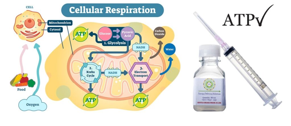
ATP(三磷酸腺苷)是人體細胞的能量分子,能啟動各種生理反應。缺乏 ATP 會導致細胞受損,甚至死亡。特別是在手術後,組織暫時缺氧,會導致 ATP 減少,影響傷口癒合與毛囊生長。
為什麼植髮後需要 ATP?
– 促進毛囊細胞修復與再生;
– 維持細胞活性,減少移植後壞死風險;
– 改善再血管化,加速恢復與生長;
– 適用於手術傷口、延遲癒合個案與植髮病人。

脂質體 ATP(Lyposomal ATP):突破性技術

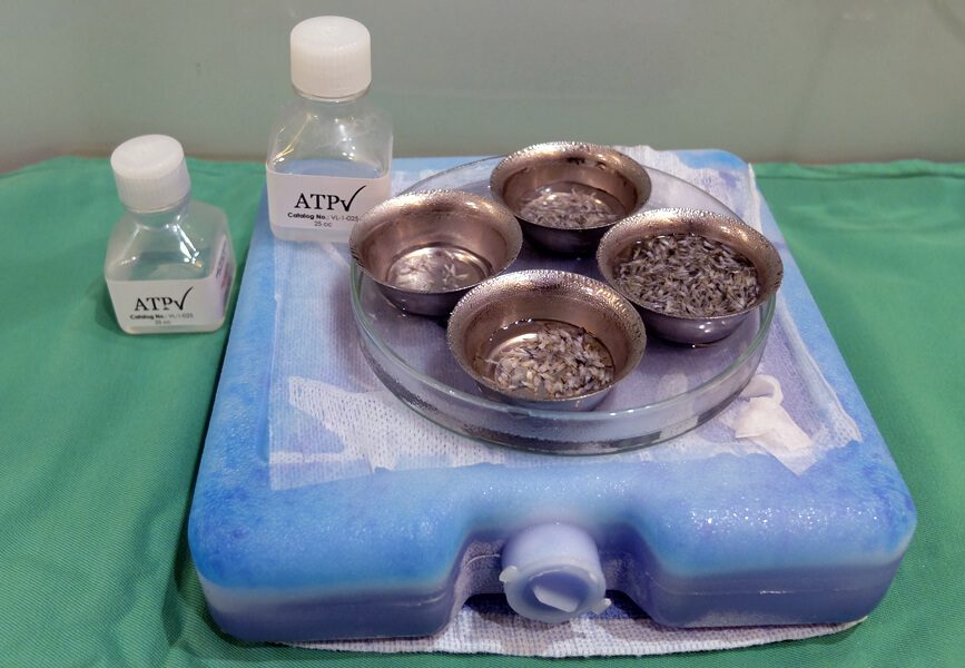
傳統 ATP 水溶液無法被細胞有效吸收 – 脂質體包裹技術(Liposome Delivery),可將 ATP 成分直接輸送進入細胞。

此項技術由美國路易斯維爾大學 William Ehringer 教授於 2002 年研發,歷經十年實驗,於 2012 年創立 Energy Delivery Solutions 公司,現已廣泛應用於術後修復、慢性傷口癒合及毛囊移植醫學。

我們在植髮手術期間與術後使用 ATP – 促進毛囊癒合、提升生長率的再生醫學技術。

國際醫學專家實證有效 : 美國 Dr. Cooley 及我們的醫療顧問 Dr. Pathomvanich 皆證實,在植髮手術中使用脂質體 ATP(Lyposomal ATP),能:
– 加速傷口癒合;
– 明顯提升毛囊成活率與生長結果。
2015 年,美絲植髮創辦人伍文輝醫生親自拜訪 ATP 發明人 Dr. Ehringer,與 Dr. Cooley 一同建議以下應用方式:

1. 手術期間:將毛囊保存在含 ATP 的保存液中;
2. 術後居家護理:前 48 小時(最多可持續 5 天)每日噴灑 ATP 配方於受區頭皮上。

臨床觀察成果: 經過一年以上的試驗觀察,結果令人驚艷,自 2016 年 10 月起,本中心為所有植髮病人常規使用脂質體 ATP;由於 ATP 半衰期極短,必須每數週從美國空運新鮮配方,我們堅持高品質,即使成本更高、過程繁瑣。
美國進口 Tricomin 銅胜肽毛囊噴霧,搭配術後護理,提升整體植髮成果。

有效成分

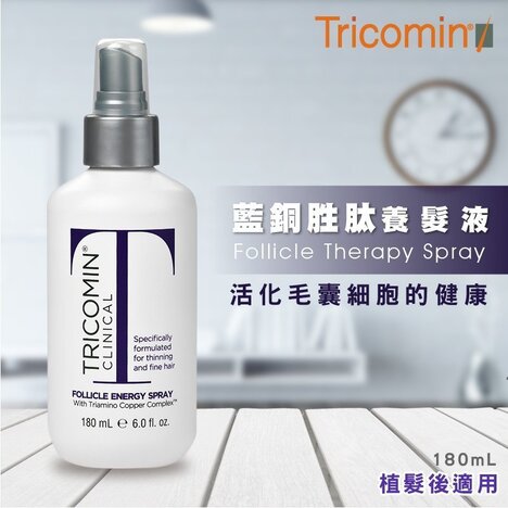
來自美國的 Tricomin Follicle Spray,含 Copper Nutritional Complex(銅營養複合物),僅使用美國原裝進口產品,確保品質穩定、安全有效。

噴霧功效

  • 促進移植毛囊健康生長;
  • 保護毛囊免受環境因素傷害,降低脫落風險;
  • 可能有助於改善脫髮根本原因。
  • Triamino Copper Nutritional Complex – 強化頭皮天然防禦機制,有效保護毛囊不受環境傷害,提升抗氧化力,為頭皮築起第一道防線。
  • Panthenol 維他命原 B5 – 滋養與修復受損髮絲,改善乾燥與毛躁。
  • 胺基酸與礦物質複合物 – 為頭髮增加豐盈感,重建髮絲強韌結構。

誰適合使用 Tricomin 毛囊噴霧?

  • 建議 所有植髮病人術後持續使用至少 4 個月;
  • 女性尤其建議使用,有助預防術後「休止期脫髮」(Shock Loss)現象;
  • 多數患者會持續使用 6–12 個月,並提早觀察到新生頭髮的萌芽與生長。

術後 4 個月內可能出現的常見反應:
– 植髮手術對頭皮產生創傷,可能導致暫時性脫髮(Shock Loss);
– 少數患者會出現移植區輕微腫脹、發紅或不適感;
– 新植入的毛囊可能在初期難以適應新環境,導致延遲生長或暫停期。

  • 減少術後脫髮(Shock Loss)
  • 強化毛囊結構,提高成活率與生長機率
  • 減輕術後腫脹、發紅與炎症反應

激光如何提供細胞能量?

激光育髮技術可活化細胞內的粒線體,促進能量產生,強化脆弱毛囊。激光進入細胞後被粒線體吸收
刺激三磷酸腺苷(ATP)的生成,這是毛囊細胞進行生長的主要能量形式。對於處於虛弱或創傷恢復期的毛囊,充足的能量供應至關重要,可促進毛囊細胞修復、增生與再生,有助於毛髮更穩定生長。

提升植髮成功率

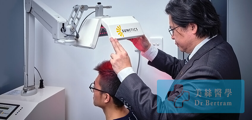
臨床認證、全球實證、有效提升毛囊存活率,全球超過數千間植髮中心(如 Bosley、HairClub)廣泛使用
美國 FDA 認證技術,現已引入本中心,專為術後使用。有別於市面廉價 LED 手持儀器,真正能深入毛囊底層,激活粒線體。

每次療程僅需 20 分鐘,喚醒細胞能量中心、打造更強韌毛囊、活化細胞內粒線體、促進 ATP 能量生成、
增強移植毛囊存活能力、提高轉化為健康終末毛髮的機率。
額外存活毛囊 = 更濃密自然的成果 : 你看不到的細節,決定最終的成敗,存活的「額外毛囊」將轉化為粗壯、自然的毛髮,提高整體密度與分佈均勻性,減少休止期脫髮,讓您更快看見成效。

什麼是「 毛囊截斷 Graft Transection 」?

術中不可忽視的風險,直接影響成效與成本:當植髮過程中使用器械角度或深度不正確,可能切斷或損傷毛囊;被截斷的毛囊失去底部幹細胞與真皮乳突,無法重新生長新髮,嚴重截斷會大幅降低植髮存活率、造成浪費。

一旦毛囊被截斷,其重新長出健康毛髮的能力將顯著降低——甚至完全喪失——導致毛囊存活率低及供區資源浪費。要將截斷率降至最低,需要精準的技術、豐富的經驗,以及在適當放大倍率下使用精細儀器。
因此,外科技巧與技術在實現持久、自然成果中扮演關鍵角色。

部分截斷後的毛囊存活率

  • 下三分之一被去除 — 毛囊存活率為 65%
  • 下二分之一被去除 — 毛囊存活率為 40%
  • 下三分之二被去除 — 無毛囊存活

過度去除幹細胞

促進毛囊再生的幹細胞存在於毛囊周圍的組織中,去除毛囊周圍過多組織也是一種物理性損傷。

Seager 在 1997 年報導:「胖毛囊」(脂肪多)比「瘦毛囊」(脂肪少)有較佳存活率。
Beehner 在 2010 年指出:差異在於胖毛囊保留了幹細胞,而瘦毛囊在修剪過程中將幹細胞去除。

我們如何減少毛囊損傷?
  • 使用 10 倍放大顯微鏡保留含幹細胞的組織 – 顯微鏡使用由 Dr. Limmer(美國)於 1991 年首次引入,並成為黃金標準;
  • FUE 採收時使用最小直徑 0.8mm 的打孔器,以保留脂肪組織中的幹細胞;
  • 使用超細手術工具;
  • 採用上述措施後,毛囊存活率可提升至超過 98%;
  • 顯微鏡技術雖於 1991 年引入,但花了 10 年才成為標準程序。

脫水會造成嚴重毛囊損傷:Gandelman:毛囊乾燥超過 5 分鐘,細胞結構開始產生不良變化;Kim:毛囊暴露於空氣超過 10 分鐘,將有超過 6% 死亡;超過 20 分鐘後,存活率急劇下降。

我們如何預防脫水?
  • 不將毛囊放置於手套手指上;
  • FUE 建議勿超過 3,000 毛囊( 約 6,000 根毛髮 );
  • 避免使用植入器(Implanter);
  • 所有毛囊皆浸泡於 ATP 溶液中。
手術時間長短如何影響毛囊存活?

插入過程可能需時 4–8 小時,經過的時間對毛髮生長與存活率至關重要,時間越長,存活率越低,隨著氧氣耗盡,二氧化碳、糖分、尿素等廢物積累,新植入的毛囊將進入酸中毒狀態。

植入後的 48–96 小時為關鍵期 – 若血液循環未能成功重建(初次血管吻合),則毛囊無法糾正代謝失衡,將導致死亡。

提取後經過的時間( 小時 )
% 毛囊生長率
2 95
4 90
6 86
8 86
24 79

從表格中可見,毛囊存活率在 4 小時後下降至 90%。換言之,超過 4 小時後,將有 10% 的提取毛囊無法生長毛髮。由於人生中的供區毛髮數量有限,這些未能成長的毛囊即為浪費。

我們如何縮短延誤時間:
  • 將 FUE 採取時間限制在 2–3 小時內;
  • 經過良好規劃的手術程序以縮短手術時間;
  • 配置大型手術團隊,分擔工作量;
  • 每天只進行一個手術案例,以專注每一位病人。

這是一種物理性損傷 – 研究指出所有冷凍的毛囊都會死亡,家用冰箱的溫度可能會波動,若溫度設定過低可能不知不覺間降至 0°C 以下,導致毛囊死亡。Kim 的研究指出:在 6 小時內,不論將毛囊儲存在室溫或 4°C,存活率無顯著差異。

我們如何預防冷凍損傷

– 若可在 4–6 小時內完成手術,將毛囊儲存於室溫;
– 使用特殊儲存液(Hypothermosol)將毛囊置於冰箱;
– 儘量於 6 小時內完成植入程序。

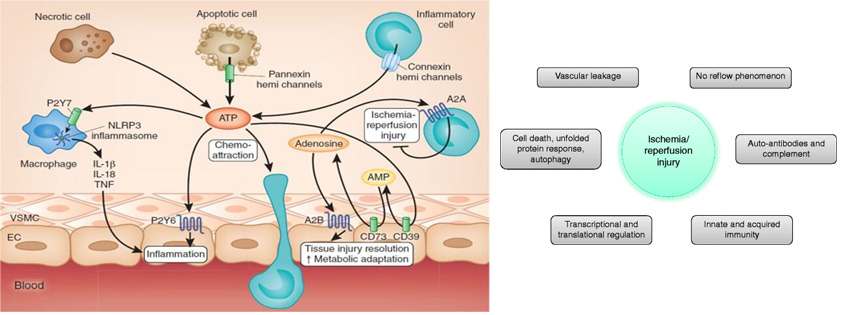
當毛囊經歷低氧環境一段時間後,在植入過程中重新暴露於氧氣中,可能會產生自由基。這些自由基可能導致細胞損傷及毛髮生長不良,這種現象稱為「缺血再灌流損傷」( Ischaemic Reperfusion Injury, IRI )。

我們如何預防缺血再灌流損傷( IRI )
  • 使用抗發炎藥物來抑制組織損傷;
  • 在儲存液中加入 ATP 以保護毛囊;
  • 縮短缺血時間,加速再血管化階段;
  • 透過激光育髮療程及減少吸煙,以維持頭皮的健康血液供應。
醫生評語

將毛囊從一個位置移植到另一個位置,並不保證一定有好結果。由於受損的毛囊無法再生,醫生必須在每一個步驟、每一個細節上都保持高度注意。

醫生全程跟進

從諮詢、手術到術後護理,您將得到醫生的持續個人護理,而不是一般顧問。

植髮前後比較

請即聯絡我們!

如有任何疑問,歡迎隨時致電本中心客服。

Whatsapp
Wechat
Instagram
LINE

國際專業認證

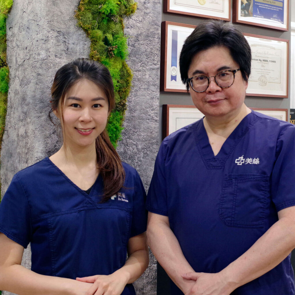
美絲是香港及中國大陸現今植髮界中最具資歷的植髮中心之一,獲多個國際醫學機構專業認可。 我們秉持最高的專業操守、為受脫髮困擾人士,提供最安全和有效的醫療解決方案。

您重拾自信之路,從這裡開始…

免費線上評估

植髮是改善髮線上移頭髮稀疏的終極解決方案,但並非每個人都適合。
我們的線上評估有助於確定這些程序是否適合您,從而節省您的求診的時間和成本。

This site is registered on wpml.org as a development site. Switch to a production site key to remove this banner.