植髮最難過的並非手術過程,而是等待頭髮生長的漫長日子。毛囊從捐髮區取出,移植到禿髮的部位,需要至少一星期才會重生接駁血管,加上植入毛囊會給頭皮造成創傷,引起腫脹,做成短暫性的缺氧。
移植的毛囊細胞為了要適應新環境,會逼使進入休止期,所以九成之上移植的毛囊髮梢,會於手術後六個星期內䬰脫落,這種暫時性的脫髮被稱為 Shock Loss,要六至八個月內再重新長出。雖然這是正常的反應,但很多病人會非常擔心,渴望頭髮會快些生長出來。經過多年的臨床實證,發現於植髮後使用低能量激光,有以下好處:
• 能加速植髮區的傷口癒合,減少手術後腫脹,發紅和發炎
• 減少結痂,通常植髮後第四天大部份的痂都已脫落
• 增加了照射部位的血液供應,減少脫髮 Shock Loss
• 有助縮短移植毛囊的休止期,頭髮更迅速地長出,提高毛囊的存活率
針對雄性禿與各類脫髮問題,雷射育髮提供細胞層級的解決方案,近年來,雷射技術已廣泛應用於多款育髮裝置中,包括:激光梳(Laser Comb)、激光帽(Laser Cap)、吹風機型激光儀(Hairdryer-like Devices)。
這些裝置可用於治療遺傳性或後天性脫髮問題,其核心原理在於雷射能量可直達毛囊細胞層級,有效活化與修復逐漸萎縮的毛囊
植髮手術在初期可能對頭皮造成暫時性創傷,常見現象包括前4個月出現暫時性脫髮(Shock Loss)。移植的供區毛囊可能需時間適應新環境 , 結合激光療程的臨床益處,包括減少術後掉髮(降低 Shock Loss 機率);
強化毛囊活力;顯著提高移植後存活率 、 和減輕術後腫脹、紅腫與發炎反應,加快恢復過程。
激光育髮技術可活化細胞內的粒線體,促進能量產生,強化脆弱毛囊。激光進入細胞後被粒線體吸收
刺激三磷酸腺苷(ATP)的生成,這是毛囊細胞進行生長的主要能量形式。對於處於虛弱或創傷恢復期的毛囊,充足的能量供應至關重要,可促進毛囊細胞修復、增生與再生,有助於毛髮更穩定生長。
手術後前4個月可能出現暫時性脫髮(Shock Loss),激光治療有以下功效:減少術後掉髮、提升移植毛囊的存活率、降低紅腫、腫脹與發炎反應、和強化術後毛囊能量供應(透過ATP刺激粒線體)。 我們採用 FDA 核准之高能量激光設備,20分鐘療程即可啟動毛囊修復能力。此舉可讓更多「存活移植株」成功轉為健康的永久毛髮。
髮源與激光醫學的歷史沿革 : 1960年代起,激光已被應用於傷口癒合研究,Finsen 博士因紫外線療法獲得 1904 諾貝爾獎,現今激光與LED光療已被廣泛應用於數萬名病患身上。
低能量激光與LED光療已被廣泛應用於多種醫療領域,激光與LED光源可直接照射於傷口或受損部位(如外傷、術後傷口),和特定身體區域(如針灸穴位、肌肉觸發點)。 臨床研究證實的應用成效,包括中風後功能恢復、加速傷口癒合、骨科疾病治療、和緩解慢性炎症。
前臨床研究顯示的潛在療效,包括脊髓損傷修復、周邊神經再生、心臟病後組織再生、神經退化性腦病(如阿茲海默症)改善、和頭部創傷(TBI)後的細胞修復。
低能量激光育髮治療, 又稱為低能量雷射治療(LLLT)、 冷雷射治療、 光調節療法(Photobiomodulation)、
生物刺激療法(Biostimulation)、光照療法(Phototherapy)… 已在數千篇經同儕審查的研究中證實,可提升細胞存活率、增殖與功能。 激光被細胞內的粒線體吸收後,會產生以下作用:
問題不再是光是否具有生物作用,而是治療性激光,和 LED 的能量,如何在細胞和生物體水平上起作用,以及這些光源的不同用途的最佳光參數是什麼。
在細胞培養、動物測試、和臨床研究中的多項研究,已證明的一個重要觀點,是相向劑量的相反作用。
LLLT 之所以稱為 「 低能量激光 」,是相對於更高頻率及功率的激光而言,當激光的光劑量低於某值,會有生髮作用相反。
光療應用方式多樣化 : 此領域的發展呈現出高度技術多元性,包括使用不同光源:激光(Laser)與LED(發光二極體); 參數變化多元,包括波長、輸出功率、連續波或脈衝模式 、脈衝頻率與時間 、和偏光狀態等 。
臨床研究與發展狀況 : 多項隨機對照試驗(Controlled Trials)顯示,光療對疼痛有顯著改善,在部分客觀指標上也觀察到進步。不過,儘管缺乏大規模支持性臨床研究,仍有數種光源已被核准作為紅外線熱療燈的替代品,用於治療各類肌肉骨骼系統疾病。
低能量激光治療(LLLT)其實早在古代已有雛形。古埃及時期,人們已利用「日光療法」作為醫療用途,這可視為人類對光線治療的最早應用。真正現代醫學上的LLLT,始於1967年。匈牙利布達佩斯賽梅維斯大學的Endre Mester教授,當時正研究激光輻射是否會引發癌症。
他剃除實驗老鼠背部毛髮,分成兩組,使用694nm波長的低能量紅寶石雷射進行照射。
實驗結果讓人驚訝:
雖然老鼠未出現癌變,但接受激光照射的老鼠,毛髮竟然長得比未照射組快!這是「雷射生物刺激」(Laser Biostimulation)首次被證實能促進毛髮生長,也標誌著 LLLT 在植髮與脫髮治療上的潛力正式誕生。
並非所有毛囊同時受到影響,進展速度也有差異
某些男性連此區亦可能逐漸稀疏
LLLT作用原理|光線如何激活細胞能量 – 紅光與近紅外光如何影響細胞?多項行動光譜(Action Spectra)分析指出,細胞色素C氧化酶(cytochrome c oxidase)是哺乳類細胞對紅光與近紅外光(Red-NIR)反應的主要光受體。儘管涉及不同生理過程,這些光譜的高峰波長幾乎一致,顯示其機制具有共通性。
為什麼細胞色素 C 氧化酶是關鍵?此酵素具備4個金屬反應中心:
– 鐵中心:haem a 及 haem a3(又稱 cytochrome a 和 a3);
– 銅中心:CuA 和 CuB。
酵素的氧化還原狀態不同,會產生不同的吸光波譜:
– 完全氧化狀態:Fe(III)、Cu(II);;
– 完全還原狀態:Fe(II)、Cu(I);;
– 亦存在多種混合價態(Mixed-Valence States);
吸收光子後,會使酵素進入激發態,加快電子傳遞反應,從而影響細胞能量代謝。
科學驗證的LLLT有效波長範圍 – 根據Karu研究團隊的最新實驗,細胞色素C氧化酶的光吸收高峰分布於以下波段:
1) 613.5 – 623.5 nm
2) 667.5 – 683.7 nm
3) 750.7 – 772.3 nm
4) 812.5 – 846.0 nm
這些波長範圍被證實最能啟動LLLT的生物效應。
光激活後的細胞反應:
– ATP合成增加:電子傳遞加快,促進粒線體產生更多ATP能量 ;
– 離子泵啟動:提升Na+/H+、Ca2+/Na+交換,活化Na+/K+ ATP酶與鈣離子幫浦 ;
– 提升第二訊息分子 ;
– ATP水平上升 → 增加cAMP生成 ;
– 鈣離子(Ca²⁺)與cAMP共同參與體內幾乎所有生理機能,包括:肌肉收縮、神經訊號傳遞、血液凝固、基因表現等。
為什麼這對植髮與脫髮治療如此關鍵?
– 促進毛囊細胞能量代謝,提高毛囊活性與再生能力;
– 加強頭皮微循環與氧氣供應,改善毛髮生長環境 ;
– 作為無創、無副作用的輔助療法,適合術前術後輔助應用。
Mester 博士首次報告:694nm 紅寶石激光可促進小鼠毛髮再生。
黑毛小鼠於第5至第7次治療後,出現明顯濃密的新生毛髮。白毛小鼠反應較慢,但亦於後期顯現效果。
第9次治療後,小鼠背部中央的毛髮生長停止,但周邊呈環狀增生現象。
對照組(未治療)小鼠的毛髮則生長緩慢或幾乎無變化。
儘管 LLLT 在市面上應用廣泛,真正符合醫學標準的人體臨床研究仍然有限:
日本研究 : 採用 Super Lizer(波長600–1600nm)治療 圓禿(Alopecia Areata), 47% 病患脫髮症狀有明顯改善。
西班牙研究 : 使用氦氖激光(HeNe laser)針對 雄性禿與圓禿治療,顯示出對不同類型脫髮均有作用。
芬蘭研究 : 比較多種光源照射頭皮後之反應,發現頭皮血流顯著增加,有助於毛囊健康與生長。
3.1 神經生長因子(NGF)與毛髮生長週期
NGF 與其受體 TrkA 在 初期生長期(anagen) 活躍。
NGF mRNA 在早期生長期達高峰,NGF 蛋白則於退化期(catagen)達高峰。
若在毛囊早期使用商用 NGF,有助於 啟動並維持生長期;然而若在後期使用,則可能促進毛囊進入退化期。
➔ 推論:NGF/TrkA 系統 對延長生長期具有正面作用。
3.2 p75 神經營養因子受體(p75NTR)與退化期控制
p75NTR 在退化期發揮促進細胞凋亡的功能。
p75NTR 缺失的小鼠,其毛囊進入退化期的時間顯著延後;而 NGF 過度表現的小鼠則提早進入退化期。
➔ p75NTR 是調控毛囊退化期的關鍵因子。
3.3 低能量激光(LLLT)提升神經生長因子(NGF)表現
HeNe 激光照射使肌肉細胞中的 NGF mRNA 上升五倍。
人類角質細胞在激光照射後,也會釋放更多 NGF。
➔ LLLT 可能透過 NGF/p75NTR 訊號通路促進毛髮再生。
4.1 肝素酶(Heparanase)
肝素酶能分解細胞外基質, 有助於幹細胞遷移與毛囊再生。
在小鼠實驗中,過度表現肝素酶加快毛髮再生,包括化療後脫髮的恢復也有顯著效果。
4.2 胸腺素β4(Thymosin Beta-4, TB4)
TB4 可促進幹細胞遷移、分化及細胞外基質重建。
在大鼠與小鼠中,能啟動毛囊幹細胞並促進毛髮生長。
4.3 Activin / Follistatin 系統
Activin 為皮膚與毛髮發育的調節因子。
Follistatin 為 Activin 的抑制蛋白,過度表現會延遲毛囊發育與進入生長週期。該系統的平衡對於髮際線重建與毛囊更新極為關鍵。
目前尚無研究直接證實 LLLT(低能量激光)對以下分子通路的作用:
Heparanase(肝素酶)
Thymosin Beta-4(TB4)
Activin / Follistatin 系統
而,這些分子已顯示與 毛囊再生、幹細胞活化與髮線調節 密切相關,被視為未來探索 LLLT 作用機轉的潛力標的。
Mester 博士的早期動物研究證明:LLLT 可有效促進動物毛髮再生
人體臨床數據雖仍有限,但呈現出明確的正向趨勢
NGF / p75NTR 訊號途徑目前被認為是最具潛力的。
Heparanase、TB4 與 Activin 等路徑,也可能在其中扮演重要角色,值得後續深入探討與臨床驗證。
超過 1,000 項研究顯示: 激光與低能量光源可提升細胞存活率、增生能力與功能活性,多項臨床隨機對照試驗已證明:LLLT 能有效刺激與保護因雄性禿,與其他脫髮病症受損的毛囊。
[1] E. Mester, B. Szende and P. Gartner, The effect of laser beams on the growth of hair in mice, Radiobiol Radiother (Berl) 9 (1968) 621-6.
[2] R. Roelandts, The history of phototherapy: something new under the sun?, J Am Acad Dermatol 46 (2002) 926-30.
[3] A.N. Pereira, P. Eduardo Cde, E. Matson and M.M. Marques, Effect of low-power laser irradiation on cell growth and procollagen synthesis of cultured fibroblasts, Lasers Surg Med 31 (2002) 263-7.
[4] J.S. Kana, G. Hutschenreiter, D. Haina and W. Waidelich, Effect of low-power density laser radiation on healing of open skin wounds in rats, Arch Surg 116 (1981) 293-6.
[5] A.P. Sommer, A.L. Pinheiro, A.R. Mester, R.P. Franke and H.T. Whelan, Biostimulatory windows in low-intensity laser activation: lasers, scanners, and NASA’s light-emitting diode array system, J Clin Laser Med Surg 19 (2001) 29-33.
[6] J.C. Sutherland, Biological effects of polychromatic light, Photochem Photobiol 76 (2002) 164-70.
[7] T. Karu, Laser biostimulation: a photobiological phenomenon, J Photochem Photobiol B 3 (1989) 638-40.
[8] T.I. Karu and N.I. Afanas’eva, Cytochrome c oxidase as the primary photoacceptor upon laser exposure of cultured cells to visible and near IR-range light, Dokl Akad Nauk 342 (1995) 693-5.
[9] R.A. Capaldi, F. Malatesta and V.M. Darley-Usmar, Structure of cytochrome c oxidase, Biochim Biophys Acta 726 (1983) 135-48.
[10] I. Szundi, G.L. Liao and O. Einarsdottir, Near-infrared time-resolved optical absorption studies of the reaction of fully reduced cytochrome c oxidase with dioxygen, Biochemistry 40 (2001) 2332-9.
[11] T.I. Karu and S.F. Kolyakov, Exact action spectra for cellular responses relevant to phototherapy, Photomed Laser Surg 23 (2005) 355-61.
[12] D. Pastore, M. Greco and S. Passarella, Specific helium-neon laser sensitivity of the purified cytochrome c oxidase, Int J Radiat Biol 76 (2000) 863-70.
[13] V.G. Artyukhov, O.V. Basharina, A.A. Pantak and L.S. Sveklo, Effect of helium-neon laser on activity and optical properties of catalase, Bull Exp Biol Med 129 (2000) 537-40.
[14] W. Yu, J.O. Naim, M. McGowan, K. Ippolito and R.J. Lanzafame, Photomodulation of oxidative metabolism and electron chain enzymes in rat liver mitochondria, Photochem Photobiol 66 (1997) 866-71.
[15] S. Passarella, He-Ne laser irradiation of isolated mitochondria, J Photochem Photobiol B 3 (1989) 642-3.
[16] S.J. Ehrreich and R.F. Furchgott, Relaxation of mammalian smooth muscles by visible and ultraviolet radiation, Nature 218 (1968) 682-4.
[17] H. Chaudhry, M. Lynch, K. Schomacker, R. Birngruber, K. Gregory and I. Kochevar, Relaxation of vascular smooth muscle induced by low-power laser radiation, Photochem Photobiol 58 (1993) 661-9.
[18] R. Mittermayr, A. Osipov, C. Piskernik, S. Haindl, P. Dungel, C. Weber, Y.A. Vladimirov, H. Redl and A.V. Kozlov, Blue laser light increases perfusion of a skin flap via release of nitric oxide from hemoglobin, Mol Med 13 (2007) 22-9.
[19] L. Vladimirov, A., G.I. Klebanov, G.G. Borisenko and A.N. Osipov, Molecular and cellular mechanisms of the low intensity laser radiation effect, Biofizika 49 (2004) 339-50.
[20] Y.A. Vladimirov, A.N. Osipov and G.I. Klebanov, Photobiological principles of therapeutic applications of laser radiation, Biochemistry (Mosc) 69 (2004) 81-90.
[21] G.G. Borisenko, A.N. Osipov, K.D. Kazarinov and A. Vladimirov Yu, Photochemical reactions of nitrosyl hemoglobin during exposure to low-power laser irradiation, Biochemistry (Mosc) 62 (1997) 661-6.
[22] B. Beltran, A. Mathur, M.R. Duchen, J.D. Erusalimsky and S. Moncada, The effect of nitric oxide on cell respiration: A key to understanding its role in cell survival or death, Proc Natl Acad Sci U S A 97 (2000) 14602-7.
[23] G.C. Brown, Regulation of mitochondrial respiration by nitric oxide inhibition of cytochrome c oxidase, Biochim Biophys Acta 1504 (2001) 46-57.
[24] N. Lane, Cell biology: power games, Nature 443 (2006) 901-3.
[25] G.C. Brown and V. Borutaite, Nitric oxide inhibition of mitochondrial respiration and its role in cell death, Free Radic Biol Med 33 (2002) 1440-50.
[26] P. Ghafourifar and E. Cadenas, Mitochondrial nitric oxide synthase, Trends Pharmacol Sci 26 (2005) 190-5.
[27] E. Clementi, G.C. Brown, N. Foxwell and S. Moncada, On the mechanism by which vascular endothelial cells regulate their oxygen consumption, Proc Natl Acad Sci U S A 96 (1999) 1559-62.
[28] F. Antunes, A. Boveris and E. Cadenas, On the mechanism and biology of cytochrome oxidase inhibition by nitric oxide, Proc Natl Acad Sci U S A 101 (2004) 16774-9.
[29] T.I. Karu, L.V. Pyatibrat and N.I. Afanasyeva, Cellular effects of low power laser therapy can be mediated by nitric oxide, Lasers Surg Med 36 (2005) 307-14.
[30] V. Borutaite, A. Budriunaite and G.C. Brown, Reversal of nitric oxide-, peroxynitrite- and S-nitrosothiol-induced inhibition of mitochondrial respiration or complex I activity by light and thiols, Biochim Biophys Acta 1459 (2000) 405-12.
[31] G.A. Guzzardella, M. Fini, P. Torricelli, G. Giavaresi and R. Giardino, Laser stimulation on bone defect healing: an in vitro study, Lasers Med Sci 17 (2002) 216-20.
[32] H. Tuby, L. Maltz and U. Oron, Modulations of VEGF and iNOS in the rat heart by low level laser therapy are associated with cardioprotection and enhanced angiogenesis, Lasers Surg Med 38 (2006) 682-8.
[33] Y. Moriyama, E.H. Moriyama, K. Blackmore, M.K. Akens and L. Lilge, In vivo study of the inflammatory modulating effects of low-level laser therapy on iNOS expression using bioluminescence imaging, Photochem Photobiol 81 (2005) 1351-5.
[34] M.C. Leung, S.C. Lo, F.K. Siu and K.F. So, Treatment of experimentally induced transient cerebral ischemia with low energy laser inhibits nitric oxide synthase activity and up-regulates the expression of transforming growth factor-beta 1, Lasers Surg Med 31 (2002) 283-8.
[35] H. Friedmann, R. Lubart, I. Laulicht and S. Rochkind, A possible explanation of laser-induced stimulation and damage of cell cultures, J Photochem Photobiol B 11 (1991) 87-91.
[36] M. Eichler, R. Lavi, A. Shainberg and R. Lubart, Flavins are source of visible-light-induced free radical formation in cells, Lasers Surg Med 37 (2005) 314-9.
[37] K. Plaetzer, T. Kiesslich, B. Krammer and P. Hammerl, Characterization of the cell death modes and the associated changes in cellular energy supply in response to AlPcS4-PDT, Photochem Photobiol Sci 1 (2002) 172-7.
[38] R. Lubart, M. Eichler, R. Lavi, H. Friedman and A. Shainberg, Low-energy laser irradiation promotes cellular redox activity, Photomed Laser Surg 23 (2005) 3-9.
[39] R. Duan, T.C. Liu, Y. Li, H. Guo and L.B. Yao, Signal transduction pathways involved in low intensity He-Ne laser-induced respiratory burst in bovine neutrophils: a potential mechanism of low intensity laser biostimulation, Lasers Surg Med 29 (2001) 174-8.
[40] F.Q. Schafer and G.R. Buettner, Redox environment of the cell as viewed through the redox state of the glutathione disulfide/glutathione couple, Free Radic Biol Med 30 (2001) 1191-212.
[41] H. Liu, R. Colavitti, Rovira, II and T. Finkel, Redox-dependent transcriptional regulation, Circ Res 97 (2005) 967-74.
[42] M. Yang, N.B. Nazhat, X. Jiang, S.M. Kelsey, D.R. Blake, A.C. Newland and C.J. Morris, Adriamycin stimulates proliferation of human lymphoblastic leukaemic cells via a mechanism of hydrogen peroxide (H2O2) production, Br J Haematol 95 (1996) 339-44.
[43] W.G. Kirlin, J. Cai, S.A. Thompson, D. Diaz, T.J. Kavanagh and D.P. Jones, Glutathione redox potential in response to differentiation and enzyme inducers, Free Radic Biol Med 27 (1999) 1208-18.
[44] S. Alaluf, H. Muir-Howie, H.L. Hu, A. Evans and M.R. Green, Atmospheric oxygen accelerates the induction of a post-mitotic phenotype in human dermal fibroblasts: the key protective role of glutathione, Differentiation 66 (2000) 147-55.
[45] T. Karu, Primary and secondary mechanisms of action of visible to near-IR radiation on cells, J Photochem Photobiol B 49 (1999) 1-17.
[46] O. Tiphlova and T. Karu, Action of low-intensity laser radiation on Escherichia coli, Crit Rev Biomed Eng 18 (1991) 387-412.
[47] T.I. Karu, L.V. Pyatibrat, G.S. Kalendo and R.O. Esenaliev, Effects of monochromatic low-intensity light and laser irradiation on adhesion of HeLa cells in vitro, Lasers Surg Med 18 (1996) 171-7.
[48] P. Moore, T.D. Ridgway, R.G. Higbee, E.W. Howard and M.D. Lucroy, Effect of wavelength on low-intensity laser irradiation-stimulated cell proliferation in vitro, Lasers Surg Med 36 (2005) 8-12.
[49] D. Hawkins and H. Abrahamse, Biological effects of helium-neon laser irradiation on normal and wounded human skin fibroblasts, Photomed Laser Surg 23 (2005) 251-9.
[50] H.S. Yu, C.S. Wu, C.L. Yu, Y.H. Kao and M.H. Chiou, Helium-neon laser irradiation stimulates migration and proliferation in melanocytes and induces repigmentation in segmental-type vitiligo, J Invest Dermatol 120 (2003) 56-64.
[51] S. Young, P. Bolton, M. Dyson, W. Harvey and C. Diamantopoulos, Macrophage responsiveness to light therapy, Lasers Surg Med 9 (1989) 497-505.
[52] Y. Fujimaki, T. Shimoyama, Q. Liu, T. Umeda, S. Nakaji and K. Sugawara, Low-level laser irradiation attenuates production of reactive oxygen species by human neutrophils, J Clin Laser Med Surg 21 (2003) 165-70.
[53] Y.S. Chen, S.F. Hsu, C.W. Chiu, J.G. Lin, C.T. Chen and C.H. Yao, Effect of low-power pulsed laser on peripheral nerve regeneration in rats, Microsurgery 25 (2005) 83-9.
[54] M. Miloro, L.E. Halkias, S. Mallery, S. Travers and R.G. Rashid, Low-level laser effect on neural regeneration in Gore-Tex tubes, Oral Surg Oral Med Oral Pathol Oral Radiol Endod 93 (2002) 27-34.
[55] P. Balaban, R. Esenaliev, T. Karu, E. Kutomkina, V. Letokhov, A. Oraevsky and N. Ovcharenko, He-Ne laser irradiation of single identified neurons, Lasers Surg Med 12 (1992) 329-37.
[56] K.R. Byrnes, R.W. Waynant, I.K. Ilev, X. Wu, L. Barna, K. Smith, R. Heckert, H. Gerst and J.J. Anders, Light promotes regeneration and functional recovery and alters the immune response after spinal cord injury, Lasers Surg Med 36 (2005) 171-85.
[57] S.O. el Sayed and M. Dyson, Effect of laser pulse repetition rate and pulse duration on mast cell number and degranulation, Lasers Surg Med 19 (1996) 433-7.
[58] R.A. Lopes-Martins, R. Albertini, P.S. Martins, J.M. Bjordal and H.C. Faria Neto, Spontaneous effects of low-level laser therapy (650 nm) in acute inflammatory mouse pleurisy induced by Carrageenan, Photomed Laser Surg 23 (2005) 377-81.
[59] A.D. Agaiby, L.R. Ghali, R. Wilson and M. Dyson, Laser modulation of angiogenic factor production by T-lymphocytes, Lasers Surg Med 26 (2000) 357-63.
[60] S. Passarella, E. Casamassima, S. Molinari, D. Pastore, E. Quagliariello, I.M. Catalano and A. Cingolani, Increase of proton electrochemical potential and ATP synthesis in rat liver mitochondria irradiated in vitro by helium-neon laser, FEBS Lett 175 (1984) 95-9.
[61] M. Greco, G. Guida, E. Perlino, E. Marra and E. Quagliariello, Increase in RNA and protein synthesis by mitochondria irradiated with helium-neon laser, Biochem Biophys Res Commun 163 (1989) 1428-34.
[62] D. Pastore, M. Greco, V.A. Petragallo and S. Passarella, Increase in H+/e- ratio of the cytochrome c oxidase reaction in mitochondria irradiated with helium-neon laser, Biochem Mol Biol Int 34 (1994) 817-26.
[63] Y. Zhang, S. Song, C.C. Fong, C.H. Tsang, Z. Yang and M. Yang, cDNA microarray analysis of gene expression profiles in human fibroblast cells irradiated with red light, J Invest Dermatol 120 (2003) 849-57.
[64] R.F. Lyons, R.P. Abergel, R.A. White, R.M. Dwyer, J.C. Castel and J. Uitto, Biostimulation of wound healing in vivo by a helium-neon laser, Ann Plast Surg 18 (1987) 47-50.
[65] H.S. Yu, K.L. Chang, C.L. Yu, J.W. Chen and G.S. Chen, Low-energy helium-neon laser irradiation stimulates interleukin-1 alpha and interleukin-8 release from cultured human keratinocytes, J Invest Dermatol 107 (1996) 593-6.
[66] V.K. Poon, L. Huang and A. Burd, Biostimulation of dermal fibroblast by sublethal Q-switched Nd:YAG 532 nm laser: collagen remodeling and pigmentation, J Photochem Photobiol B 81 (2005) 1-8.
[67] N. Kipshidze, V. Nikolaychik, M.H. Keelan, L.R. Shankar, A. Khanna, R. Kornowski, M. Leon and J. Moses, Low-power helium: neon laser irradiation enhances production of vascular endothelial growth factor and promotes growth of endothelial cells in vitro, Lasers Surg Med 28 (2001) 355-64.
[68] A. Khanna, L.R. Shankar, M.H. Keelan, R. Kornowski, M. Leon, J. Moses and N. Kipshidze, Augmentation of the expression of proangiogenic genes in cardiomyocytes with low dose laser irradiation in vitro, Cardiovasc Radiat Med 1 (1999) 265-9.
[69] A.R. Medrado, L.S. Pugliese, S.R. Reis and Z.A. Andrade, Influence of low level laser therapy on wound healing and its biological action upon myofibroblasts, Lasers Surg Med 32 (2003) 239-44.
[70] E.J. Neiburger, Rapid healing of gingival incisions by the helium-neon diode laser, J Mass Dent Soc 48 (1999) 8-13, 40.
[71] J.T. Eells, M.M. Henry, P. Summerfelt, M.T. Wong-Riley, E.V. Buchmann, M. Kane, N.T. Whelan and H.T. Whelan, Therapeutic photobiomodulation for methanol-induced retinal toxicity, Proc Natl Acad Sci U S A 100 (2003) 3439-44.
[72] M.T. Wong-Riley, H.L. Liang, J.T. Eells, B. Chance, M.M. Henry, E. Buchmann, M. Kane and H.T. Whelan, Photobiomodulation directly benefits primary neurons functionally inactivated by toxins: role of cytochrome c oxidase, J Biol Chem 280 (2005) 4761-71.
[73] D. Gigo-Benato, S. Geuna and S. Rochkind, Phototherapy for enhancing peripheral nerve repair: a review of the literature, Muscle Nerve 31 (2005) 694-701.
[74] J.J. Anders, S. Geuna and S. Rochkind, Phototherapy promotes regeneration and functional recovery of injured peripheral nerve, Neurol Res 26 (2004) 233-9.
[75] J.J. Anders, R.C. Borke, S.K. Woolery and W.P. Van de Merwe, Low power laser irradiation alters the rate of regeneration of the rat facial nerve, Lasers Surg Med 13 (1993) 72-82.
[76] K. Branco and M.A. Naeser, Carpal tunnel syndrome: clinical outcome after low-level laser acupuncture, microamps transcutaneous electrical nerve stimulation, and other alternative therapies–an open protocol study, J Altern Complement Med 5 (1999) 5-26.
[77] J. Irvine, S.L. Chong, N. Amirjani and K.M. Chan, Double-blind randomized controlled trial of low-level laser therapy in carpal tunnel syndrome, Muscle Nerve 30 (2004) 182-7.
[78] M.I. Weintraub, Noninvasive laser neurolysis in carpal tunnel syndrome, Muscle Nerve 20 (1997) 1029-31.
[79] Y. Ueda and N. Shimizu, Pulse irradiation of low-power laser stimulates bone nodule formation, J Oral Sci 43 (2001) 55-60.
[80] Y. Ueda and N. Shimizu, Effects of pulse frequency of low-level laser therapy (LLLT) on bone nodule formation in rat calvarial cells, J Clin Laser Med Surg 21 (2003) 271-7.
[81] M.S. Ribeiro, F. Da Silva Dde, C.E. De Araujo, S.F. De Oliveira, C.M. Pelegrini, T.M. Zorn and D.M. Zezell, Effects of low-intensity polarized visible laser radiation on skin burns: a light microscopy study, J Clin Laser Med Surg 22 (2004) 59-66.
[82] T. Moshkovska and J. Mayberry, It is time to test low level laser therapy in Great Britain, Postgrad Med J 81 (2005) 436-41.
[83] L.A. Santana-Blank, E. Rodriguez-Santana and K.E. Santana-Rodriguez, Photo-infrared pulsed bio-modulation (PIPBM): a novel mechanism for the enhancement of physiologically reparative responses, Photomed Laser Surg 23 (2005) 416-24.
[84] M.L. Kripke, Ultraviolet radiation and immunology: something new under the sun–presidential address, Cancer Res 54 (1994) 6102-5.
[85] P. Whittaker, Laser acupuncture: past, present, and future, Lasers Med Sci 19 (2004) 69-80.
雷射與LED:對脫髮的作用有何不同?
雖然市面上常見的 低能量光療(LLLT) 裝置包含 雷射與LED兩種類型,但兩者在生髮效果與原理上並不完全相同。
射(Laser):具備集中穿透力,效果更一致。雷射光為相干光(coherent light),能量集中且具方向性,可深入滲透至頭皮深層,有效刺激毛囊生長;
通常搭配優化波長(如650–850nm),臨床成效更穩定,常應用於雄性禿、女性頭髮稀疏、髮際線後移治療。
LED:光線較分散,穿透較淺,效果因人而異 ; 屬於非相干光(non-coherent light),發散且不具方向性
穿透力較淺,對毛囊的刺激效果較為有限,部分用於家用儀器或輔助保養,但臨床數據變異較大。
結論:選擇光源類型影響治療成效 : 雖然 雷射與LED都可促進毛髮生長,但在 臨床應用中,雷射裝置通常帶來更穩定且可預測的效果。建議在選擇 低能量光療法(LLLT) 時, 優先考慮專業雷射設備與波長匹配性,提升療效。


國際專業認證
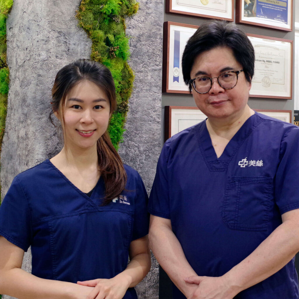
美絲是香港及中國大陸現今植髮界中最具資歷的植髮中心之一,獲多個國際醫學機構專業認可。 我們秉持最高的專業操守、為受脫髮困擾人士,提供最安全和有效的醫療解決方案。
植髮是改善髮線上移頭髮稀疏的終極解決方案,但並非每個人都適合。
我們的線上評估有助於確定這些程序是否適合您,從而節省您的求診的時間和成本。