What Kills Your Grafts ?

 How Surgical Skill & Aftercare Prevent Damage to Irreplaceable Follicles

20 Years of Expertise : Every Step Performed in Hong Kong By Doctors

What can damage or kill hair transplant grafts?

A hair transplant’s success is measured by graft survival—the percentage of transplanted follicles that grow permanently. Grafts are living tissues that can be damaged or killed by human error (H-Factors) and biological stressors (X-Factors). Once a graft is damaged, it cannot regenerate. Protecting grafts requires meticulous surgical technique, strict sterile protocols, and precise patient aftercare at every stage.

Grafts can be damaged or killed by two main categories: “H-Factors” (human/protocol error) and “X-Factors” (biological stressors). These can occur during harvesting, storage, implantation, or healing. Because the total number of lifetime donor follicles is fixed and irreplaceable, maximizing survival is the paramount goal of ethical hair restoration.

Sara’s Comment

After a person is born, there will not be an additional hair follicle. Every step of the procedure should be carefully designed to maximize graft survival, ensuring each transplanted follicle has the best chance to grow strong and last a lifetime.

Content-index

What Are "X-Factors" (Biological Stressors)?

X-Factors are subclinical biological injuries that are not always visible but compromise graft viability. They now account for the majority of unexplained poor growth.

  • Reperfusion Injury: Oxidative damage when blood supply is re-established to the graft.

  • Overheating & Dehydration: In the first critical week post-op, before new circulation is secure.

  • Micro-inflammation: An exaggerated or prolonged inflammatory response that damages the follicle.

  • Auto-rejection: A rare immunological response to the transplanted tissue.

What Are "H-Factors" (Human/Protocol Errors)?

H-Factors represent preventable damage caused by a breach in surgical protocol or patient non-compliance.

H-Factors Before Procedure

  • Poor Recipient Site Circulation: Scarring or vascular conditions that impair blood flow to the transplanted area.

  • Uncontrolled Health Conditions: Smoking, uncontrolled diabetes, or nutritional deficiencies that compromise healing.

H-Factors During Procedure (Most Critical Phase)

  • Dehydration: Grafts exposed to air without proper hydration.

  • Physical Trauma: Crushing, transecting (cutting), or excessive handling of follicles.

  • Improper Storage: Use of non-physiological solutions or temperature extremes (cold injury).

  • Prolonged Out-of-Body Time: Grafts left outside the body for too long before implantation.

H-Factors After Procedure

  • Patient Non-Compliance: Failure to follow post-op instructions (e.g., scratching, sun exposure, strenuous activity).

  • Physical Trauma: Direct impact to the recipient area.

  • Infection: Poor wound care leading to folliculitis.

Why is Graft Survival the Most Important Metric?

  • Finite Donor Supply: You have a limited, non-renewable bank of permanent donor follicles. Wastage is permanent loss.

  • Directly Impacts Final Result: Survival rate dictates density. Studies suggest at least 85% survival is needed for a good cosmetic outcome.

  • Affects Future Sessions & Scarring: High survival in the first session preserves more donor hair for future needs and can allow for a smaller donor scar (in FUT).

  • Determines True Cost: Clinics charge per graft transplanted, not per graft grown. A low survival rate drastically increases your real cost per surviving graft.

Comparative Analysis: The Hidden Cost of Poor Survival
  • Center A (20% Damage Rate): Paying $31 per surviving graft.

  • Center B (2% Damage Rate): Paying $25.50 per surviving graft.

  • Conclusion: A clinic with lower technical skill (higher transection/damage rate) is more expensive in reality, as you pay for grafts that never grow.

How Does a Healthy Graft Heal and Revascularize?

Understanding the biology underscores why technique is critical.

  • Day 1-3: Graft is in ischemic phase, surviving on diffusion. Inflammation begins.

  • Day 3-7: Critical period for revascularization (new blood vessel formation). Neutrophils and macrophages are active.

  • Week 2-4: Inflammatory cells subside; fibroblasts and collagen stabilize the graft.

  • Month 1-3: Follicle enters new growth cycle, often shedding the original hair shaft before producing new hair.

How Do We Maximize Graft Survival?

Our protocol is designed to systematically minimize H and X Factors.

  1. Pre-Op Optimization: Medical screening and patient guidance to improve scalp health.

  2. Surgical Precision: Use of high-magnification microscopes, chilled physiological storage solutions, and refined instruments to minimize trauma and dehydration.

  3. Trained, Coordinated Team: More skilled hands reduce out-of-body time. We aim for large sessions to be efficient, not rushed.

  4. Detailed Aftercare Plan: Specific instructions on washing, activity, and monitoring to protect grafts during the fragile healing phase.

Key Takeaways:

  • Grafts Are Irreplaceable: Damaged hair follicles cannot be regenerated or cloned. Survival is the cornerstone of ethical surgery.

  • Two Killers: H-Factors & X-Factors: Failure results from human/protocol error (H) or biological injury (X). Expert teams work to minimize both.

  • Survival Dictates True Value: The real cost is calculated per surviving graft. A “cheaper” clinic with high transection rates is often more expensive long-term.

  • The First Week is Critical: The most vulnerable period is during surgery and the immediate post-op days before revascularization is complete.

  • Skill Over Speed: A methodical, precise surgical team using optimal protocols achieves higher survival than a fast, high-volume assembly line.

Your Next Step: Ask About Our Survival Protocols

Choosing a surgeon is choosing a custodian for your finite donor hair. Ask our doctors during your consultation about our specific graft survival protocols, storage solutions, and average out-of-body times. We are transparent about the measures we take to protect your most valuable asset—your donor follicles.

Graft survival rates vary by individual and procedure. Adherence to all pre- and post-operative instructions is critical. This information is for educational purposes and details a specific surgical philosophy.

References

  • Aby Mathew. A review of Cellular Bippreservation Consideration During Hair Transplantation. Hair Transplant Forum International, Vol 23 No. 1, January/February 2013
  • Baust, J.G. In: J.G. Baust and J.M. Baust, eds. Advances in Biopreservation. CRC Press, 2007; 1-14.
  • Snyder, K.K., et al. Biological packaging for the global cell and tissue therapy markets. BioProcessing Journal. 2004(May/June); 1-7.
  • Reyes, A.B., J.S. Pendergast, and S. Yamazaki. Mammalian peripheral circadian oscillators are temperature compensated. J Biol Rhythms. 2008; 23:95-98.
  • Allen, F.M. Physical and toxic factors in shock. Archives of Surgery. 1939; 38:155-180.
  • Bigelow, W.G., W.K. Lindsay, and W.F. Greenwood. Hypothermia: its possible role in cardiac surgery: an investigation of factors governing survival in dogs at low body temperature. Annals of Surgery. 1950; 132:849-866.
  • Swan, H., et al. Hypothermia in surgery: analysis of 100 clinical cases. Annals of Surgery. 1955; 142:382-400.
  • Collins, G.M., M. Bravo-Shugarman, and Pl. Terasaki. Kidney preservation for transplantation: initial perfusion and 30 hours ice storage. Lancet. 1969; 2:1219.
  • Taylor, M.J., et al. A new solution for life without blood: asanguineous low flow perfusion of a whole-body perfusate during 3 hours of cardiac arrest and profound hypothermia. Circulation. 1995; 91:431-444.
  • Van Buskirk, R.G., et al. Assessment of hypothermic storage of normal human epidermal keratinocytes (NHEK) using Alamar Blue. In Vitro Toxicology. 1996; 9:297-303.
  • Mathew, A.J., J.G. Baust, and R.G. Van Buskirk. Optimization of HypoThermosol for the hypothermic storage of cardiac cells—addition of EDTA. In Vitro Toxicology. 1997; 10(4):407-415.
  • Mathew, A.J., et al. Vitamin E and EDTA improve the efficacy of HypoThermosol—implication of apoptosis. In Vitro Toxicology. 1999; 12(3):163-172.
  • Dahdah, N.S., et al. Effects of HypoThermosol, an experimental acellular solution for tissue preservation and cardiopulmonary bypass, on isolated newborn lamb coronary vessels subjected to ultraprofound hypothermia and anoxia. Cryobiology. 1999; 39:58-68.
  • Mathew, A.J., J.G. Baust, and R.G. Van Buskirk. Improved hypothermic preservation of human renal cells through suppression of both apoptosis and necrosis. Cell Preservation Technology. 2003; 1:239-254.
  • Baust, J.M., et al. Transplantation diagnostics: utilization of protein microarray analysis to determine kidney status and transplantation efficacy. Cell Preservation Technology. 2004; 2(2):81.
  • Mathew, A.J., et al. Cell preservation in reparative and regenerative medicine: evolution of individualized solution composition. Tissue Engineering. 2004; 10:1662-1671.
  • Snyder, K.K., et al. Biological packaging for the global cell and tissue therapy markets. BioProcessing Journal. 2004; 3(3):39.
  • Van Buskirk, R.G., et al. Hypothermic storage and cryopreservation: the issues of successful short-term and long term preservation of cells and tissues. BioProcess Int’l. 2004; 2(10):42.
  • Baust, J.M. Advances in media for cryopreservation and storage. BioProcess International. 2005; 3:46-56.
  • Snyder, K.K., et al. Enhanced hypothermic storage of neonatal cardiomyocytes. Cell Preservation Technology. 2005; 3(1):61-74.
  • Mathew, A.J. I’m losing cell viability and function at different points in my process, and I don’t know why! BioProcess Int’l. 2010; 8(6) 54-7.
  • Ikonomovic, M., et al. Ultraprofound cerebral hypothermia and blood substitution with an acellular synthetic solution maintains neuronal viability in rat hippocampus. CryoLetters. 2001; 22:19-26.
  • Lodish, H.F., et al. Transport across cell membranes. In: S. Tenney, ed. Molecular Cell Biology. 4th Edition. New York: W.H. Freeman, 2000; 590-591.
  • MacKnight, A.D.C., and A. Leaf. Regulation of cellular volume. Physiology Review. 1977; 57:510.
  • Martin, D.R., et al. Primary cause of unsuccessful liver and heart preservation: cold sensitivity of the ATPase system. Annals of Surgery. 1972; 175:11.
  • Boutilier, R.G. Mechanisms of cell survival in hypoxia and hypothermia. J of Experimental Biology. 2001; 204:3171-3181.
  • Toledo-Pereyra, L.H., A.J. Paez-Rollys, and J.M. PalmaVargas. Science of organ preservation. In: L.H. ToledoPereyra, ed. Organ Procurement and Preservation for Transplantation. Landes Bioscience, 1997; 1-16.
  • Rehncrona, S., B.K. Siesjo, and D.S. Smith. Reversible ischemia of the brain: biochemical factors influencing restitution. ActaPhysioliogy Scandinavia. 1979; Suppl 492:135.
  • Cotterill, P. ISHRS survey majority using chilled saline. W. Reed Personal communication, December 2012. Reed, W. Personal communication, December 2012.
  • Perez-Meza, D., M. Leavitt, and M. Mayer. The growth factors. Part 1: clinical and histological evaluation of the wound healing and revascularization of the hair graft after hair transplant surgery. Hair Transplant Forum Int’l. 2007; 17(5):173.
  • Perez-Meza D. Wound healing and revascularization of the hair transplant graft: role of the growth factors. In: W. Unger and R. Shapiro, eds. Hair Transplantation, 4th Ed. Marcel Dekker, New York, 2004; 287-294.
  • Cooley, J. Ischemia-reperfusion injury and graft storage solutions. Hair Transplant Forum Int’l. 2004; 14(4):121,127,130.
  • Parsley, W.M., and D. Perez-Meza. Review of factors affecting the growth and survival of follicular grafts. J Cutan Aesthet Surg. 2010; 3(2):69-75.
  • Limmer, R. Micrograft survival. In: D. Stough, ed. Hair Replacement. Mosby Press, 1996; 147-149.
  • Kim, J.C., and S. Hwang. The effects of dehydration, preservation temperature and time, and hydrogen peroxide on hair grafts. In: W.P. Unger and R. Shapiro, eds. Hair Transplantation, 4th Ed. New York: Marcel Dekker, 2004; 285-286.
  • Hwang, S.J., et al. The effects of dehydration, preservation temperature and time on the hair grafts. Annals of Dermatology. 2002; 14:149-152. 37. Krugluger, W., et al. Enhancement of in vitro hair shaft elongation in follicles stored in buffers that prevent follicle cell apoptosis. Dermatol Surg. 2004; 30:1-5.
  • Krugluger, W., et al. New storage buffers for micrografts enhance graft survival and clinical outcome in hair restoration surgery. Hair Transplant Forum Int’l. 2003; 13(3):333-334.
  • Van Buskirk, R.G., et al. Navigating the post-preservation viability fog: assay standardization for cell and tissue therapy applications. Genetic Engineering News. 2006; 26(19):38-39.
  • Van Buskirk, R. Viability and functional assays used to assess preservation efficacy: the multiple endpoint/tier approach. In: J.G. Baust and J.M. Baust, eds. Advances in Biopreservation. CRC Press-Taylor and Francis Publishing: New York, 2006.
  • Belzer, F., and J. Southard. Principles of solid-organ preservation by cold storage. Transplantation. 1988; 45(4):673-676.
  • Taylor, M.J. The role of pH and buffer capacity in the recovery of function of smooth muscle cooled to –13°C in unfrozen media. Cryobiology. 1982; 19:585-60.
  • Taylor, M.J., and Y. Pignat. Practical acid dissociation constants, temperature coefficients and buffer capacities for some biological buffers in solutions containing dimethyl sulfoxide between 25 and –12°C. Cryobiology. 1982; 19:99-109.
  • Fujita, J. Cold shock response in mammalian cells. J Mol Microbiol Biotechnol. 1999; 1(2):243-255.
  • Plesnila, N., et al. Effect of hypothermia on the volume of rat glial cells. Journal of Physiology. 2000; 523.1:155-162.
  • Tseng, Y-C, et al. Exploring uncoupling proteins and antioxidant mechanisms under acute cold exposure in brains of fish. PLoS ONE. 2011; 6(3):1-15.
  • Corwin, W.L., et al. The unfolded protein response in human corneal endothelial cells following hypothermic storage: Implications of a novel stress pathway. Cryobiology. 2011; 63(1):46-55.
  • Baust, J.M. Overview of hypothermic storage. Hair Transplant Forum Int’l. 2006; 16(2):53.
  • Cosentino, L.M., et al. Preliminary report: evaluation of storage conditions and cryococktails during peripheral blood mononuclear cell cryopreservation. Cell Preservation Technology. 2007; 5:189-204.
  • Baust, J.M., R.G. Van Buskirk, and J.G. Baust. Cell viability improves following inhibition of cryopreservation-induced apoptosis. In Vitro Cell and Developmental Biology. 2000; 36:262.
  • Baust, J.M., et al. A molecular basis of cryopreservation failure and its modulation to improve cell survival. Cell Transplantation. 2001; 10:561.
  • Baust, J.M., R.G. Van Buskirk, and J.G. Baust. Gene activation of the apoptotic caspase cascade following cryogenic storage. Cell Preservation Technology. 2002; 1:63.
    •   Baust, J.M. Molecular mechanisms of cellular demise associated with cryopreservation failure. Cell Preservation Technology. 2002; 1:17.
  • Anderson, R.V., M.G. Siegman, and R.S. Balaban. Hyperglycemia increases cerebral intracellular acidosis during circulatory arrest. Annals of Thoracic Surgery. 1992; 54:1126-1130.
  • Ely, S.W., and R.M. Berne. Protective effects of adenosine in myocardial ischemia. Circulation. 1992; 85:893-904.
  • Bessems, M., et al. Preservation of rat livers by cold storage: a comparison between the University of Wisconsin solution and HypoThermosol. Ann Transplant. 2004; 9(2):35-37.
  • Povsic, T., et al. A double-blind, randomized, controlled, multicenter study to assess the safety and cardiovascular effects of skeletal myoblast implantation by catheter delivery in patients with chronic heart failure after myocardial infarction. American Heart Journal. 2011; 162(4):654-662.
  • Powell, R., et al. Interim analysis results from the RESTORE-CLI, a randomized, double-blind multicenter phase II trial comparing expanded autologous bone marrow-derived tissue repair cells and placebo in patients with critical limb ischemia. J of Vasc Surg.2011; 54(4):1032-1041.
  • Ginis, I., B. Grinblat, and M. Shirvan. Evaluation of bone marrow-derived mesenchymal stem cells after cryopreservation and hypothermic storage in clinically safe medium. Tissue Engineering Part C Methods. 2012; 18(6):453-463.
  • Lowe, N.J., P.L. Lowe, and J. St Clair Roberts. A phase IIa open-label dose-escalation pilot study using allogeneic human dermal fibroblasts for nasolabial folds. Dermatol Surg. 2010; 36(10):1578-1585.
  • Raposio, E., et al. Effects of cooling micrografts in hair transplantation surgery. Dermatol Surg. 1999; 25:705-707.
    •   Jiange, Q., et al. How long can hair follicle units be preserved at 0 and 4°C for delayed transplant? Dermatol Surg. 2005; 31:23-26.
  • Kurata, S., et al. Viability of isolated single hair follicles preserved at 4°C. Dermatol Surg. 1999; 25:26-29.
  • Beehner, M.L. Notes from the Editor Emeritus. Hair Transplant Forum Int’l. 2005; 15(6):193-195.
  • Cooley, J.E. Successful extended storage of hair follicles using hypothermic media with liposomal ATP. Translational Regenerative Medicine Forum. 2010.
  • Beehner, M.L. 96-hour study of FU graft “out-of-body” survival comparing saline to HypoThermosol/ATP solution. Hair Transplant Forum Int’l. 2011; 21(2):1, 37.
  • Ehringer, W.D. New horizons in storage solutions and additive agents in organ transplantation. Norwood Lecture, ISHRS 2011 Annual Scientific Meeting.
36

MEDICAL - DISCLAIMERS

Images & Information shown are for reference only

Information on this website is provided for general educational purposes only and does not constitute personalized medical advice. It is not intended to promote our service or imply superiority over another.

Individual results in hair restoration vary significantly and no outcome can be guaranteed. The before-and-after images shown represent possible results — not promises. We recommend seeking independent medical advice to discuss your options … Read More

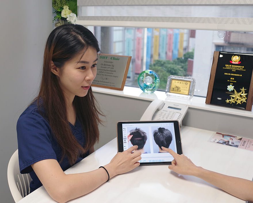
Pre-Consultation Review

Ensure You're a Good Candidate for hair transplant

Why pay for a consultation if you’re not a candidate? Start with our free WhatsApp assessment — the first step in our ‘No-Regret’ protocol to see if hair transplant is a realistic solution for your hair loss.

Still Have Questions ?

If you want to get a personalized answer from our medical team, you can reach us using this form here. We will cantact you as soon as possible.

Our Quality & Safety Standards

Our practice adheres to guidelines established by leading international organizations in Hair Restoration.

ISHRS

International Society of Hair Restoration Surgery is the leading global medical association that establishes international practice standards and patient safety protocols.

ABHRS

The American Board of Hair restoration Surgery represents the highest standard. To maintain rigorous certification requirements the physician must demonstrate surgical expertise.

WFI

Worls FUE InstituteI serves as the premier educational body focused exclusively on Follicular Unit Extraction methodology. The institute ensures consistent application of safe FUE.

This site is registered on wpml.org as a development site. Switch to a production site key to remove this banner.