Graft Survival

What Kills Your Transplanted Grafts?

Transplanting a graft from one site to another does not guarantee good result. As damaged graft cannot re-grow, the surgeon must pay attention to every step and every details of the procedure. On average 85-92% of grafts survive after transplant and grow new hair.

2 possible ways a graft can be damaged or even killed – the X-Killing Factor and the H-Killing Factor.

X Graft Killing Factors

“X” stands for unknown. The term was first described by Dr Shield (Australia) in 1984, when the reduced hair growth was unexpected and unexplained. In early years this X-Factor only accounts for a very small 0.5-1% of all no growth. With the advance and standardization of surgicial techniques, X-Factors now accounts for the majority of unsatisfactory results.

Possible X Factors

It is of our opionion that such “unknown” factors are in fact biological damage to the grafts which takes plce during and after the procedure, including:

  • Denied or missed H-Factors
  • Reperfusion Ischaemic Injury
  • Auto-rejection of Grafts
  • Overheating and Dehydration of Grafts in the First Week
  • Improper haircare or activities

“H” stands for human. Poor growth is caused by a breach of protocol. H-factor may occur before, during, and after the procedure. The result of a hair transplant procedure cannot therefore be guaranteed.

H-Factors Before Procedure

Predisposing Factors That Compromise Result include:

  • Poor Circulation of the Recipient Area
  • Scar on to-be-transplanted areas
  • Smoking
  • Uncontrolled Diabetes
H- Factors During Procedure

Grafts are injured During Procedure:

  • Dehydration
  • Excessive Removal of Supporting Tissue
  • Cold Injury
  • Transecting the Hair Follicles
  • Chemical Trauma To Grafts During Storage
  • Ischaemic-Reperfusion Injury
H-Factors After Procedure

Transplanted Follicles are injured after the procedure

  • Failure To Compile With Instruction
  • Failure to Form New Circulation
  • Physical Trauma To Transplanted Graft
  • Infection

A hair follicle is a living tissue. A lot of things take place after the follicle is planted into the new site. Understanding of the natural histochemical process is crucial, as a better graft survival means a better outcome.

After the graft is harvested from the donor area, it is placed in a storage solution for preservation until implanted in the recipient area. Its original blood supply is detached. Oxygen is depleted and waste products begin to accumulate. The ability of the graft to receive nutrients from the recipient site and undergo revascularization (formation of new blood vessels) therefore determines survival.

After the graft is inserted into one of the slits, the wound healing process immediately takes place. There is a cascade of cellular activities – edema ( swelling ), erythema ( redness ), hair shedding, new hair formation, and the revascularization of the hair graft.

Histological Changes

  • On day 1, only edema is found
  • By day 3, important influx of neutrophils and occasional eosinophils
  • By day 7, neutrophils and macrophages are both present
  • By day 14, predominant cell types seen are macrophages, fibroblasts, lymphocytes, occasional histiocytes
  • From days 21 to 28, inflammatory cells decrease and then disappear
  • Collagen III and IV present from day 1 to 6 months

Quick Navigation

Graft Survival Affects Hair Transplant Result

At Dr. Bertram Hair Transplant Centre, our approach is built around one core principle: quality matters more than quantity. Every step of our procedure is carefully designed to maximize graft survival, ensuring that each transplanted follicle has the best chance to grow strong and last a lifetime.

After a person is born, there will not be an additional hair follicle. The permanent hair follicles at the back of our head, the donor site, are invaluable and cannot be replaced. Many people expect hair follicles to be cloned, but the date of success is an unknown. The priority is to minimize the trauma of hair follicles during the process of hair transplantation.

Over the past 50 years, many studies have been conducted to look at the actual survival rate of transplanted hair, and the percentage growth, reporting the following findings :

  • Hair is considered an “organ” for transplantation
  • Like other organs, they must be carefully stored after leaving the body, otherwise they will die
  • Damaged hair follicles may not produce new hair after transplantation
  • Generally requires at least 85% of transplanted hair to grow to see result
  • When more grafts survive, less is required to complete the job.
  • As width of the FUT donor scar is proportional to the width of the strip excised, a higher survival will reduce the donor scar.

The final result of the hair transplant procedure depends on how many grafts actually regrow. Dr Beehner commented that the percentage of graft survival depends on the skill of the surgical team more than anything. In another word, the skill and experience of the surgical team is one of the most important factors in arriving at satisfactory result.

The doctor and the staff should not just focus on extraction, but also on how to keep it growing after transplanting into another site. Taking out a graft cannot guarantee its growth unless the H-Factors and X-Factors are looked into. We therefore saw the importance to improve graft survival.

  • Hair transplant is challenged by the limited donor supply.
  • Until now Hair Cloning is still not available.
  • The best way to ensure adequate supply of hair follicles for the next procedure is to minimize graft wastage in the first session.

When more grafts survive, less is required to complete the job. As most hair transplant centers charge “per graft transplanted” not “per graft survives”, a higher survival will bring the cost down. Having say that, money cannot buy back a wastage of good donor hair follicles.

 
Center A
Center B
Cost per Graft (HKD) 25 25
No of Graft Required 2,400 2,400
Total Cost (HKD) 60,000 60,000
Transection Rate (Graft damage) 20% 2%
No. of Damage Graft 2,400 X 20% = 480 2,400 X 2% = 48
No. of Intact Graft 2,400 - 480 = 1,920 2,400 - 48 = 2,352
Actual Cost per Graft (HKD) 60,000 ÷ 1,920 = 31 60,000 ÷ 2,352 = 25.5

CONCLUSION : A high transection rate increases the cost per graft.

  • Aby Mathew. A review of Cellular Bippreservation Consideration During Hair Transplantation. Hair Transplant Forum International, Vol 23 No. 1, January/February 2013
  • Baust, J.G. In: J.G. Baust and J.M. Baust, eds. Advances in Biopreservation. CRC Press, 2007; 1-14.
  • Snyder, K.K., et al. Biological packaging for the global cell and tissue therapy markets. BioProcessing Journal. 2004(May/June); 1-7.
  • Reyes, A.B., J.S. Pendergast, and S. Yamazaki. Mammalian peripheral circadian oscillators are temperature compensated. J Biol Rhythms. 2008; 23:95-98.
  • Allen, F.M. Physical and toxic factors in shock. Archives of Surgery. 1939; 38:155-180.
  • Bigelow, W.G., W.K. Lindsay, and W.F. Greenwood. Hypothermia: its possible role in cardiac surgery: an investigation of factors governing survival in dogs at low body temperature. Annals of Surgery. 1950; 132:849-866.
  • Swan, H., et al. Hypothermia in surgery: analysis of 100 clinical cases. Annals of Surgery. 1955; 142:382-400.
  • Collins, G.M., M. Bravo-Shugarman, and Pl. Terasaki. Kidney preservation for transplantation: initial perfusion and 30 hours ice storage. Lancet. 1969; 2:1219.
  • Taylor, M.J., et al. A new solution for life without blood: asanguineous low flow perfusion of a whole-body perfusate during 3 hours of cardiac arrest and profound hypothermia. Circulation. 1995; 91:431-444.
  • Van Buskirk, R.G., et al. Assessment of hypothermic storage of normal human epidermal keratinocytes (NHEK) using Alamar Blue. In Vitro Toxicology. 1996; 9:297-303.
  • Mathew, A.J., J.G. Baust, and R.G. Van Buskirk. Optimization of HypoThermosol for the hypothermic storage of cardiac cells—addition of EDTA. In Vitro Toxicology. 1997; 10(4):407-415.
  • Mathew, A.J., et al. Vitamin E and EDTA improve the efficacy of HypoThermosol—implication of apoptosis. In Vitro Toxicology. 1999; 12(3):163-172.
  • Dahdah, N.S., et al. Effects of HypoThermosol, an experimental acellular solution for tissue preservation and cardiopulmonary bypass, on isolated newborn lamb coronary vessels subjected to ultraprofound hypothermia and anoxia. Cryobiology. 1999; 39:58-68.
  • Mathew, A.J., J.G. Baust, and R.G. Van Buskirk. Improved hypothermic preservation of human renal cells through suppression of both apoptosis and necrosis. Cell Preservation Technology. 2003; 1:239-254.
  • Baust, J.M., et al. Transplantation diagnostics: utilization of protein microarray analysis to determine kidney status and transplantation efficacy. Cell Preservation Technology. 2004; 2(2):81.
  • Mathew, A.J., et al. Cell preservation in reparative and regenerative medicine: evolution of individualized solution composition. Tissue Engineering. 2004; 10:1662-1671.
  • Snyder, K.K., et al. Biological packaging for the global cell and tissue therapy markets. BioProcessing Journal. 2004; 3(3):39.
  • Van Buskirk, R.G., et al. Hypothermic storage and cryopreservation: the issues of successful short-term and long term preservation of cells and tissues. BioProcess Int’l. 2004; 2(10):42.
  • Baust, J.M. Advances in media for cryopreservation and storage. BioProcess International. 2005; 3:46-56.
  • Snyder, K.K., et al. Enhanced hypothermic storage of neonatal cardiomyocytes. Cell Preservation Technology. 2005; 3(1):61-74.
  • Mathew, A.J. I’m losing cell viability and function at different points in my process, and I don’t know why! BioProcess Int’l. 2010; 8(6) 54-7.
  • Ikonomovic, M., et al. Ultraprofound cerebral hypothermia and blood substitution with an acellular synthetic solution maintains neuronal viability in rat hippocampus. CryoLetters. 2001; 22:19-26.
  • Lodish, H.F., et al. Transport across cell membranes. In: S. Tenney, ed. Molecular Cell Biology. 4th Edition. New York: W.H. Freeman, 2000; 590-591.
  • MacKnight, A.D.C., and A. Leaf. Regulation of cellular volume. Physiology Review. 1977; 57:510.
  • Martin, D.R., et al. Primary cause of unsuccessful liver and heart preservation: cold sensitivity of the ATPase system. Annals of Surgery. 1972; 175:11.
  • Boutilier, R.G. Mechanisms of cell survival in hypoxia and hypothermia. J of Experimental Biology. 2001; 204:3171-3181.
  • Toledo-Pereyra, L.H., A.J. Paez-Rollys, and J.M. PalmaVargas. Science of organ preservation. In: L.H. ToledoPereyra, ed. Organ Procurement and Preservation for Transplantation. Landes Bioscience, 1997; 1-16.
  • Rehncrona, S., B.K. Siesjo, and D.S. Smith. Reversible ischemia of the brain: biochemical factors influencing restitution. ActaPhysioliogy Scandinavia. 1979; Suppl 492:135.
  • Cotterill, P. ISHRS survey majority using chilled saline. W. Reed Personal communication, December 2012. Reed, W. Personal communication, December 2012.
  • Perez-Meza, D., M. Leavitt, and M. Mayer. The growth factors. Part 1: clinical and histological evaluation of the wound healing and revascularization of the hair graft after hair transplant surgery. Hair Transplant Forum Int’l. 2007; 17(5):173.
  • Perez-Meza D. Wound healing and revascularization of the hair transplant graft: role of the growth factors. In: W. Unger and R. Shapiro, eds. Hair Transplantation, 4th Ed. Marcel Dekker, New York, 2004; 287-294.
  • Cooley, J. Ischemia-reperfusion injury and graft storage solutions. Hair Transplant Forum Int’l. 2004; 14(4):121,127,130.
  • Parsley, W.M., and D. Perez-Meza. Review of factors affecting the growth and survival of follicular grafts. J Cutan Aesthet Surg. 2010; 3(2):69-75.
  • Limmer, R. Micrograft survival. In: D. Stough, ed. Hair Replacement. Mosby Press, 1996; 147-149.
  • Kim, J.C., and S. Hwang. The effects of dehydration, preservation temperature and time, and hydrogen peroxide on hair grafts. In: W.P. Unger and R. Shapiro, eds. Hair Transplantation, 4th Ed. New York: Marcel Dekker, 2004; 285-286.
  • Hwang, S.J., et al. The effects of dehydration, preservation temperature and time on the hair grafts. Annals of Dermatology. 2002; 14:149-152. 37. Krugluger, W., et al. Enhancement of in vitro hair shaft elongation in follicles stored in buffers that prevent follicle cell apoptosis. Dermatol Surg. 2004; 30:1-5.
  • Krugluger, W., et al. New storage buffers for micrografts enhance graft survival and clinical outcome in hair restoration surgery. Hair Transplant Forum Int’l. 2003; 13(3):333-334.
  • Van Buskirk, R.G., et al. Navigating the post-preservation viability fog: assay standardization for cell and tissue therapy applications. Genetic Engineering News. 2006; 26(19):38-39.
  • Van Buskirk, R. Viability and functional assays used to assess preservation efficacy: the multiple endpoint/tier approach. In: J.G. Baust and J.M. Baust, eds. Advances in Biopreservation. CRC Press-Taylor and Francis Publishing: New York, 2006.
  • Belzer, F., and J. Southard. Principles of solid-organ preservation by cold storage. Transplantation. 1988; 45(4):673-676.
  • Taylor, M.J. The role of pH and buffer capacity in the recovery of function of smooth muscle cooled to –13°C in unfrozen media. Cryobiology. 1982; 19:585-60.
  • Taylor, M.J., and Y. Pignat. Practical acid dissociation constants, temperature coefficients and buffer capacities for some biological buffers in solutions containing dimethyl sulfoxide between 25 and –12°C. Cryobiology. 1982; 19:99-109.
  • Fujita, J. Cold shock response in mammalian cells. J Mol Microbiol Biotechnol. 1999; 1(2):243-255.
  • Plesnila, N., et al. Effect of hypothermia on the volume of rat glial cells. Journal of Physiology. 2000; 523.1:155-162.
  • Tseng, Y-C, et al. Exploring uncoupling proteins and antioxidant mechanisms under acute cold exposure in brains of fish. PLoS ONE. 2011; 6(3):1-15.
  • Corwin, W.L., et al. The unfolded protein response in human corneal endothelial cells following hypothermic storage: Implications of a novel stress pathway. Cryobiology. 2011; 63(1):46-55.
  • Baust, J.M. Overview of hypothermic storage. Hair Transplant Forum Int’l. 2006; 16(2):53.
  • Cosentino, L.M., et al. Preliminary report: evaluation of storage conditions and cryococktails during peripheral blood mononuclear cell cryopreservation. Cell Preservation Technology. 2007; 5:189-204.
  • Baust, J.M., R.G. Van Buskirk, and J.G. Baust. Cell viability improves following inhibition of cryopreservation-induced apoptosis. In Vitro Cell and Developmental Biology. 2000; 36:262.
  • Baust, J.M., et al. A molecular basis of cryopreservation failure and its modulation to improve cell survival. Cell Transplantation. 2001; 10:561.
  • Baust, J.M., R.G. Van Buskirk, and J.G. Baust. Gene activation of the apoptotic caspase cascade following cryogenic storage. Cell Preservation Technology. 2002; 1:63.
    •   Baust, J.M. Molecular mechanisms of cellular demise associated with cryopreservation failure. Cell Preservation Technology. 2002; 1:17.
  • Anderson, R.V., M.G. Siegman, and R.S. Balaban. Hyperglycemia increases cerebral intracellular acidosis during circulatory arrest. Annals of Thoracic Surgery. 1992; 54:1126-1130.
  • Ely, S.W., and R.M. Berne. Protective effects of adenosine in myocardial ischemia. Circulation. 1992; 85:893-904.
  • Bessems, M., et al. Preservation of rat livers by cold storage: a comparison between the University of Wisconsin solution and HypoThermosol. Ann Transplant. 2004; 9(2):35-37.
  • Povsic, T., et al. A double-blind, randomized, controlled, multicenter study to assess the safety and cardiovascular effects of skeletal myoblast implantation by catheter delivery in patients with chronic heart failure after myocardial infarction. American Heart Journal. 2011; 162(4):654-662.
  • Powell, R., et al. Interim analysis results from the RESTORE-CLI, a randomized, double-blind multicenter phase II trial comparing expanded autologous bone marrow-derived tissue repair cells and placebo in patients with critical limb ischemia. J of Vasc Surg.2011; 54(4):1032-1041.
  • Ginis, I., B. Grinblat, and M. Shirvan. Evaluation of bone marrow-derived mesenchymal stem cells after cryopreservation and hypothermic storage in clinically safe medium. Tissue Engineering Part C Methods. 2012; 18(6):453-463.
  • Lowe, N.J., P.L. Lowe, and J. St Clair Roberts. A phase IIa open-label dose-escalation pilot study using allogeneic human dermal fibroblasts for nasolabial folds. Dermatol Surg. 2010; 36(10):1578-1585.
  • Raposio, E., et al. Effects of cooling micrografts in hair transplantation surgery. Dermatol Surg. 1999; 25:705-707.
    •   Jiange, Q., et al. How long can hair follicle units be preserved at 0 and 4°C for delayed transplant? Dermatol Surg. 2005; 31:23-26.
  • Kurata, S., et al. Viability of isolated single hair follicles preserved at 4°C. Dermatol Surg. 1999; 25:26-29.
  • Beehner, M.L. Notes from the Editor Emeritus. Hair Transplant Forum Int’l. 2005; 15(6):193-195.
  • Cooley, J.E. Successful extended storage of hair follicles using hypothermic media with liposomal ATP. Translational Regenerative Medicine Forum. 2010.
  • Beehner, M.L. 96-hour study of FU graft “out-of-body” survival comparing saline to HypoThermosol/ATP solution. Hair Transplant Forum Int’l. 2011; 21(2):1, 37.
  • Ehringer, W.D. New horizons in storage solutions and additive agents in organ transplantation. Norwood Lecture, ISHRS 2011 Annual Scientific Meeting.

How We Improve Your Graft Survival

Warning : Damaged Graft Cannot Re-grow

What is ATP ?

Since 2018, we import the ATP Solution from USA

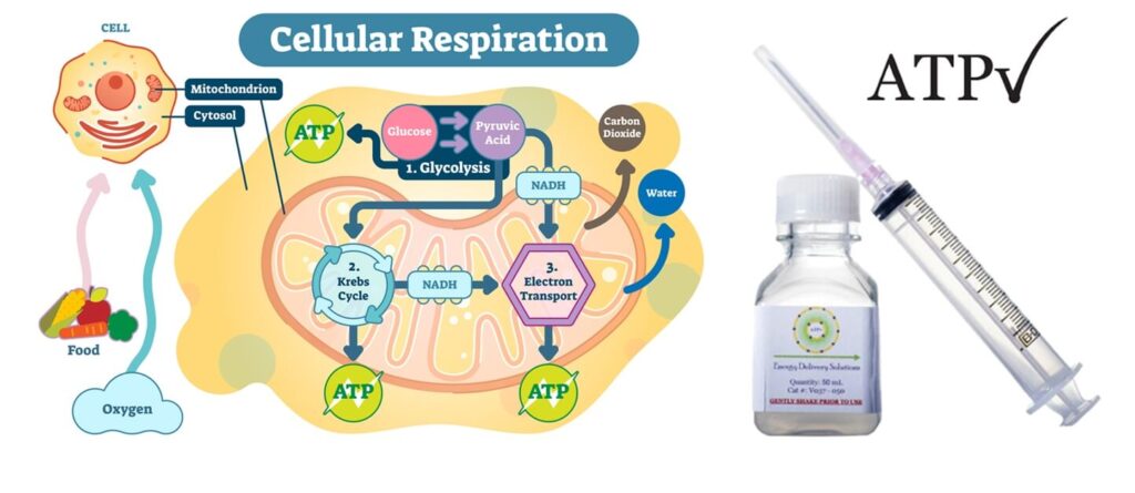
ATP (Adenosine triphosphate) is an energy molecule that catalyzes a variety of important cellular reactions in our bodies. The lack of ATP can cause cell damage and eventual cell and tissue death. Because the creation and regulation of ATP requires oxygen, tissues deprived of oxygen often experience ATP loss and become damaged. Because of this, ATP becomes crucial for patients healing from wounds; especially surgical wounds.

Lyposomal ATP – the Breakthrough

The usual kind of ATP solution in a vial is very cheap but the use is limited, as aqueous solution cannot be absorbed into cells. 

In 2002, Dr. William Ehringer a Professor at the University of Louisville, School of Medicine, discovered a method to encapsulate and deliver ATP to cells. After 10 years of research the final formulations were identified – Lyposomal ATP. In 2012, Dr. Ehringer founded Energy Delivery Solutions in manufacturing ATP for use in post-surgical patients, patients with delayed wound healing, and in hair restoration.

We Use ATP During and After Hair Transplant

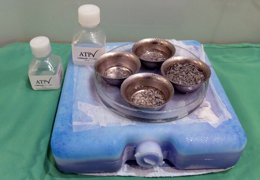
Experiments by Dr Cooley and Dr Pathomvanich our Medical Advisor showed that, in hair transplant surgery, Lyposomal ATP allows for quicker healing and better hair transplant yields. In 2015 Dr Bertram Ng met with Dr Ehringer founder of Energy Delivery Solutions. Dr Ehringer and Dr Cooley. Both recommended:

1. Store grafts in ATP-containing storage solution
2. Spray ATP Formulation at home onto the recipient scalp first 48 hours, up to 5 days

Clinical trials on selected subjects for over a year revealed very impressive result. Since October 2016 we provide Lyposomal ATP to all patients routinely. As Lyposomal ATP has only a very short half life, we have to import from the state every few weeks. The benefit is well worth the extra efforts and expenses.

Useful Constituents

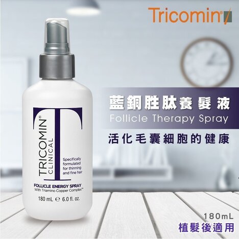
The Tricomin Follicle Spray from USA which contains Copper Nutritional Complex for use after hair transplant. The preparations we are using are fully imported from USA to ensure quality.

Benefits

  • Encourages healthy growth of the transplanted hair follicles
  • Protects transplanted grafts against environmental factors that may cause hair to fall-out more rapidly
  • May help to treat the underlying cause of hair loss
  • Triamino Copper Nutritional Complex powers-up natural defense process protect hair from environmental damage
  • Panthenol repairs and nourishes damaged hair
  • Amino acids and minerals build body in the hair to produce a fuller appearance

Who Should Consider using Tricomin Follicle Spray ?

  • We recommend all patients to use for at least 4 months after the procedure
  • Women would find it usefuk to prevent “Shock Loss” – temporary shedding of transplanted as well as existing hair
  • Most use it up to 12 months and experience an early growth of transplanted hair

Hair transplants can initially traumatize the scalp and can result in a temporary hair loss during the first 4 months (this is known as shock loss). Some patients may experience swelling in the transplanted area. The transplanted donor follicles can also experience difficulty adapting to their new environment. Clinical studies have demonstrated the following beneficial effects of laser when use in conjunction with hair transplant.

  • minimizes hair shedding (shock loss)
  • strengthen hair follicles after surgery with a much higher probability of survival
  • reduce swelling, redness and inflammation post-surgery

Effect of Laser in Cell Energy Supply

Laser hair therapy stimulates the mitochondria in cells to increase the production of adenosine triphosphate (ATP). ATP is the form of energy used by hair cells to grow imto follicles. Abundant energy supply is critical when dealing with weakened and traumatized hair follicles.

Increase Success Rate after Hair Transplant

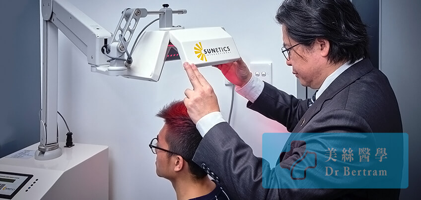
Laser hair therapy devices have been used by thousands of hair transplant centers all over the world (such as Bosley and HairClub). However handheld contraptions made with cheap Light Emitting Diodes (LEDs) are worthless when it comes to energizing the base of hair follicles. Technologically advanced device with the FDA-cleared is now available in our center for use after hair transplant.

A 20 minutes of treatment is able to revive the mitochondria of hair cells. This can result in stronger hair follicles with a higher probability of surviving the operation. These extra amounts of “survivor” hair grafts will eventually grow into healthy, terminal hairs.

Graft Transection

Graft transection occurs when a hair follicle is accidentally cut or damaged during the harvesting or implantation process in a hair transplant. This can happen if the angle or depth of the instrument used is incorrect, severing the follicle’s lower portion, which contains essential stem cells and the dermal papilla.

Once a follicle is transected, its ability to regrow healthy hair is significantly reduced – or lost entirely – leading to poor graft survival and wasted donor resources. Minimizing transection requires precision, experience, and the use of fine instruments under proper magnification. That’s why surgical skill and technique play a critical role in achieving lasting, natural-looking results.

Graft Survival After Partial Transection

  • Lower 1/3 Removed – 65% Graft Survives 
  • Lower 1/2 Removed – 40% Graft Survives
  • Lower 2/3 Removed – None Survives

Excessive Removal of Stem Cells

Removing too much tissue around the hair follicle is also a form of physical injury. Stem cells responsible for follicular re-growth is located at the tissue surrounding the graft.

Seager reported in 1997 that Chubby Graft ( more fat ) survives better than Skinny Graft ( less fat ). In 2010 Beehner commented that the differences in survival is that the stem cells are retained in chubby grafts but trimmed away in the skinny grafts

How We Reduce Graft Damage
  • Use 10X manification microscopes to preserve stem cell containing tissues. The use of microscope was first introduced by Dr Limmer (USA) in 1991 and becomes the gold standard. 
  • Use a minimal 0.8mm diameter punch in FUE for harvesting to retain the stem cells in fatty tissues
  • Use of extra-fine instruments
  • With above measures graft survival can be increased to more than 98%
  • The use of microscope was first introduced by Dr Limmer (USA) in 1991. However it took 10 years to become the gold standard. 

Dehydration causes serious graft damage. Deleterious changes in cell integrity when follicles are left to dry for more than 5 minutes (Gandelman). When grafts are exposed to air for more than 10 minutes, more than 6% will die. After 20 minutes survival markedly decreases (Kim)

How We Overcome Dehydration
  • Do not leave grafts on the gloved fingers
  • Avoid using FUE for more than 3,000 grafts ( 6,000 Hairs )
  • Avoid using implanters
  • All grafts are submerged in ATP solution
How Duration of Surgery Affects Graft Survival ?

It may take 4–8 hours to complete the insertion. The elapsed time is critical for hair growth and survival because increased time results in decreased survival. As oxygen is depleted and waste products such as carbon dioxide, sugar, and urea begin to accumulate, a newly planted hair graft will be acidotic.

The first 48–96 hours after implantation is critical. If the circulation cannot be re-established (the primary inosculation) to correct the metabolic imbalances, the hair graft won’t survive and could die.

Duration After Extraction (hour)
% of Growth
2 95
4 90
6 86
8 86
24 79

From the table you can see that graft survival to 90% after 4 hours. So after 4 hours 10% of the extracted grafts will not grow hair. This is a wastage as the donor hair is limited in a life time.

How We Shortens The Delay
  • Limited FUE Extraction time to 2-3 hours
  • Well planned procedure to shorten the operating time
  • A large surgical team to share the work load
  • Only perform one case a day so that all attention will be given

This is a form of physical injury. Study had shown that all frozen graft will die. Domestic-grade refrigerator may have temperature fluctuation. If the fridge temperature is set too low it may drop below 0°C and kill grafts without knowing. From studies there was no significant difference in survival within 6 hours, whether the grafts were stored in room temperature or 4 °C (Kim)

How We Overcome Cold Injury

•   Store graft at room temperature if procedure can be complated within 4 6 hours
•   Use a special storage solution ( Hypothermosol ) to store grafts in the fridge
•   Try to complete implantation within 6 hours

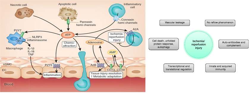
After a period of low oxygen, when the grafts are implanted and suddenly re-exposed to oxygen, they may form free radicals. These free radicals may cause cell injury and suboptimal growth, and is known as “Ischaemic Reperfusion Injury or IRI” on Re-implanted Graft.

How We Overcomes IRI
  • Suppress tissue injury by the use of anti-inflammatory medication
  • ATP is added to the storage solution to protect the grafts
  • Decrease the ischemia time to accelerate the revascularization phase.
  • Maintaining a healthy blood supply to the scalp by using Laser hair Therapy & reduce smoking
Doctor’s Comment

Transplanting a graft from one site to another does not guarantee good result. As damaged graft cannot re-grow, the surgeon must pay attention to every step and every details of the procedure. 

Doctor Care at Every Step

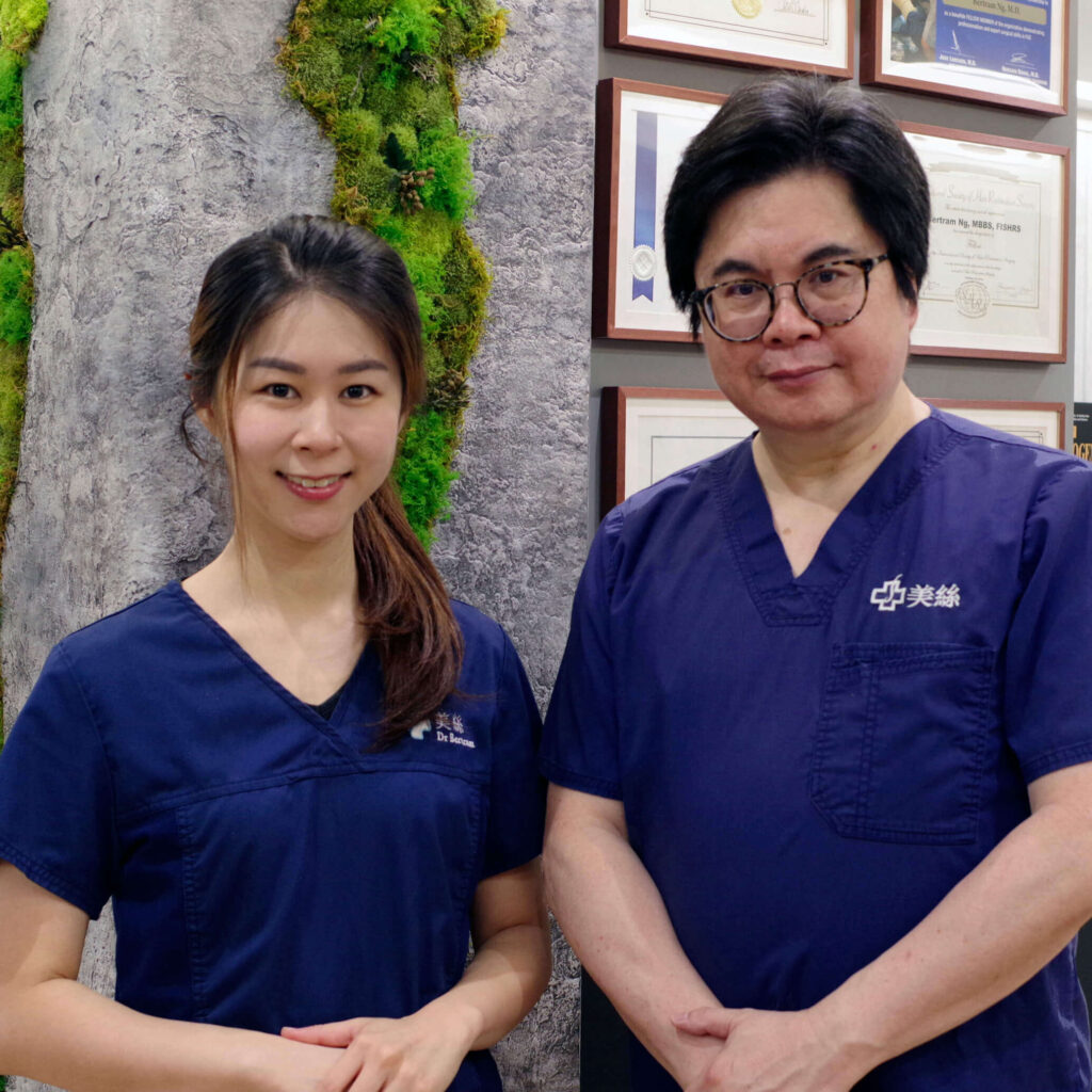
From consultation, surgery, to aftercare, you will receive continued personal care by our doctors, not just consultants.

Before & After

Contact Us Now !

Have a question? Please feel free to call our friendly customer service.

Whatsapp
Wechat
Instagram
LINE

International Accreditations

Recognized by leading global medical bodies, our clinic stands as one of the most qualified and internationally accredited hair transplant centers in Hong Kong and mainland China. We are proud to uphold the highest standards in medical ethics, safety, and surgical expertise.

Your Hair Restoration Starts Here ...

Free Online Assessment

Hair transplant is the ultimate solution to restores hair, but not everyone is good candidate. 
Our online assessment helps determine if these procedures suit you, saving you time and costs.

This site is registered on wpml.org as a development site. Switch to a production site key to remove this banner.