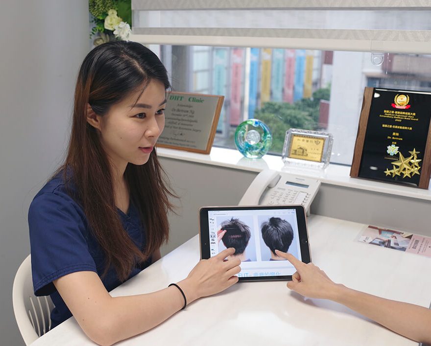
Low Level Laser Therapy

Post-Transplant Laser Therapy : Clinically Useful or Just a Marketing Gimmick?

Use of Laser in Post-Transplant Care : Our Clinical Perspective

Last Updated: 6 Nov 2025

Low-Level Laser Therapy ( LLLT ), also known as photobiomodulation, is one of only three FDA-cleared treatment modalities for androgenetic alopecia, alongside minoxidil and finasteride ( FDA, 2007; Avci et al., 2014 ). At our clinic, we have incorporated LLLT into post-operative care for over 15 years, primarily during the first 3–7 days following hair transplantation.

Our primary objective is not long-term hair growth stimulation, but rather to support early wound healing, reduce post-operative inflammation, and potentially improve graft survival during the critical initial phase when follicles are most vulnerable ( Gupta & Daigle, 2014; Lanzafame et al., 2014 ).

While some studies suggest LLLT may modestly enhance hair density with consistent use — typically requiring treatment every other day for at least 20 minutes per session ( as noted by laser therapy experts, including those from the UK’s Hair Restoration Advisory Group ) — the effect size is generally small compared to pharmacologic therapies ( Jimenez et al., 2014; Wikramanayake et al., 2020 ).

Importantly, sustained benefits for hair growth require ongoing, regular use. For this reason, we believe home-use LLLT devices ( e.g., FDA-cleared laser helmets or combs ) are more practical and cost-effective than in-office sessions, which are often too infrequent to produce meaningful regrowth effects.

Medical therapy should be grounded in evidence-based medicine, not promoted as a marketing-driven add-on. While many clinics now routinely offer post-transplant laser therapy, its benefits — particularly beyond the early healing phase — are modest at best and require consistent, long-term use to be meaningful. When offered solely as an in-clinic service without a clear protocol or patient commitment to ongoing treatment, it often becomes a time-consuming intervention with little tangible return. True value lies in scientifically supported, patient-centered care — not in ancillary services that prioritize profit over proven outcomes.

In summary :

  • Post-op ( Days 1–7 ) : LLLT may aid healing and reduce edema/swelling.
  • Long-term : Any hair growth benefit is minor and dose-dependent, best achieved via consistent home use, not sporadic clinic visits.
  • Individual results vary : LLLT is considered adjunctive — not a replacement — for medical or surgical hair restoration.


References :

  • FDA. (2007). Clearance of HairMax LaserComb for androgenetic alopecia. [K063427]
  • Avci, P., et al. ( 2014 ). Low-level laser ( light ) therapy ( LLLT ) in skin: stimulating, healing, restoring. Seminars in Cutaneous Medicine and Surgery, 33(1), 48–53.
  • Lanzafame, R. J., et al. ( 2014 ). Hair growth in men and women using a low-level laser therapy device: A multicenter, randomized, sham device–controlled, double-blind study. Lasers in Surgery and Medicine, 46(2), 144–151.
  • Jimenez, J. J., et al. ( 2014 ). Efficacy and safety of a low-level laser device in the treatment of male and female pattern hair loss: A multicenter, randomized, sham device–controlled, double-blind study. American Journal of Clinical Dermatology, 15(2), 115–127.
  • Wikramanayake, T. C., et al. ( 2020 ). Effects of low-level laser therapy on hair regrowth: A systematic review. Journal of Cosmetic and Laser Therapy, 22(5), 273–279.

Content-index

Clinical application of Laser

For Hair Loss

Laser is able to stimulate and preserve hair follicles in patients with androgenetic alopecia and other hair loss disorders. Laser has been used over the past few years in a number of laser devices ( combs, caps, hairdryer-like ) for treatment of genetic or acquired hair loss. The Laser energy addresses hair loss at the hair follicle cellular level, rejuvenating miniaturizing hair follicles in seven major ways :

  • Photo-Biomodulation and a stimulation effect
  • Initiates protein synthesis
  • Mobilizes calcium ions within the hair follicle
  • Mobilizes cellular stimulation within the dermal papilla
  • Enhancement of ATP production in the cells
  • Significant improvement of blood microcirculation
  • Allows greater nutrient acquisition by follicular site

After Hair Transplant

Hair transplants can initially traumatize the scalp and can result in a temporary hair loss during the first 4 months (this is known as shock loss). Some patients may experience swelling in the transplanted area. The transplanted donor follicles can also experience difficulty adapting to their new environment. Clinical studies have demonstrated the following beneficial effects of laser when use in conjunction with hair transplant.

  • minimizes hair shedding ( shock loss )
  • strengthen hair follicles after surgery with a much higher probability of survival
  • reduce swelling, redness and inflammation post-surgery
Advantage 1 : Effect of Laser in Cell Energy Supply
  • Laser hair therapy stimulates the mitochondria in cells to increase the production of adenosine triphosphate (ATP).
  • ATP is the form of energy used by hair cells to grow imto follicles.
  • Abundant energy supply is critical when dealing with weakened and traumatized hair follicles.
Advantage 2 : Increase Success Rate after Hair Transplant
  • Laser hair therapy devices have been used by thousands of hair transplant centers all over the world ( such as Bosley and HairClub ).
  • However handheld contraptions made with cheap Light Emitting Diodes ( LEDs ) are worthless when it comes to energizing the base of hair follicles.
  • Technologically advanced device with the FDA-cleared is now available in our center for use after hair transplant. A 20 minutes of treatment is able to revive the mitochondria of hair cells.
  • This can result in stronger hair follicles with a higher probability of surviving the operation.
  • These extra amounts of “survivor” hair grafts will eventually grow into healthy, terminal hairs.

Promotes Wound Healing

  • Since the first experiments in the 1960s with laser treatment for impaired wound healing, there has been much research-both lab-based and clinically-into the use of lasers for medical treatment.
  • Later Nils Finsen pioneered the use of UV therapy for which he won the Nobel prize in 1904 [2]).
  • The use of lasers and LEDs as light sources was the next step in the technological development of light therapy, which is now applied to many thousands of people worldwide each day.
  • The Lasers and LEDs are applied directly to the respective areas ( e.g., wounds, sites of injuries ) or to various points on the body (acupuncture points, muscle-trigger points).
  • Controlled clinical trials have shown efficacy in treating stroke, stimulating wound healing, orthopedic conditions and relief of chronic inflammation.
  • Preclinical studies have shown effectiveness in spinal cord injuries, peripheral nerve regeneration, heart attacks, degenerative brain diseases and traumatic brain injury.

Other Applications

  • Sports-medicine – reduce swelling and hematoma, relieve pain, treat acute soft-tissue injuries
  • Rehabilitation – improve mobility
  • Dentistry – treat inflamed oral tissues and heal diverse ulcerations
  • Dermatology – treat edema, non-healing ulcers, burns, and dermatitis
  • Rheumatology – relieve pain and treat chronic inflammations and autoimmune diseases
  • Veterinary Medicine – especially in racehorse-training centers
  • Other specialists and general practitioners

BioMechanism of Use in Hair Loss

What is Laser Hair Therapy ?

Laser Therapy, also referred to as Low Level Laser Therapy ( LLLT ), cold laser therapy, photobiomodulation, biostimulation, and phototherapy, has been shown in thousands of peer-reviewed publications to increase cellular survival, proliferation and function. The laser light after absorbed by mitochondria in the cell produces the following actions

  • Photo-Biomodulation and a stimulation effect
  • Initiates protein synthesis
  • Mobilizes calcium ions
  • Enhancement of ATP production
  • Significant improvement of blood microcirculation
  • Allows greater nutrient acquisition
  • Increases cellular respiration
  • Induces activation of transcription factors via reactive oxygen species.


The question is no longer whether light has biological effects but rather how energy from therapeutic lasers and LEDs works at the cellular and organism levels and what the optimal light parameters are for different uses of these light sources.

Biphasic Dose Response - Kills Hair vs Promotes Hair Growth

  • One important point that has been demonstrated by multiple studies in cell culture, animal models and in clinical studies is the concept of a biphasic dose response with the total delivered light energy density ( fluence ).
  • The reason why the technique is termed Low-level is that there exists an optimal dose of light for any particular application, and dose lower than this optimum value, or more significantly, larger than the optimum value will have a diminished therapeutic outcome, or for high doses of light a negative outcome may result.

Delivering Methods

  • The methods for delivering the therapeutic light are diverse. The field is characterized by a variety of methodologies and uses of various light sources ( lasers, LEDs ) with different parameters ( wavelength, output power, continuous-wave or pulsed operation modes, pulse parameters, polarization state etc ).
  • In 2002 MicroLight Corp received 510K FDA clearance for the ML 830-nm diode laser for treatment of carpal tunnel syndrome.
  • There were several controlled trials reporting significant improvement in pain and some improvement in objective outcome measures.
  • Since then several light sources have been approved as equivalent to an infra-red heating lamp for treating a wide-range of musculoskeletal disorders with no supporting clinical studies.

Historical Development of LLLT

1967

  • Low Level Light therapy  (LLLT ) is one of the oldest therapeutic methods used by humans ( historically as solar therapy by Egyptians ).
  • In 1967 a few years after the first working laser was invented, Endre Mester in Semmelweis University, Budapest, Hungary decided to test if laser radiation might cause cancer in mice.
  • He shaved the hair off their backs, divided them into two groups and gave a laser treatment with a low powered ruby laser ( 694-nm ) to one group.
  • They did not get cancer and to his surprise the hair on the treated group grew back more quickly than the untreated group.
  • This was the first demonstration of “laser biostimulation” to re-grow hair.

1989

  • Increase the number of the miniaturized follicles causes baldness. Hair follicles in hairline, midscalp, crown, and temples are most sensitive to DHT.
  • In men the first appearance is therefore a receding hairline and/or thinning crown.
  • Thinning eventually progresses into other areas.
  • In the more advanced AGA only a rim or “horseshoe” pattern of hair remains.
  • In some men even this remaining rim of hair can be affected by DHT.

1995

  • An analysis of five action spectra suggested that the primary photoacceptor for the red-NIR range in mammalian cells is cytochrome c oxidase [6].
  • It is remarkable that the action spectra that were analyzed had very close (within the confidence limits) peak positions in spite of the fact that these are seemingly different processes.
  • The enzyme contains two iron centres, haem a and haem a3 ( also referred to as cytochromes a and a3 ), and two copper centres, CuA and CuB [7].
  • Fully oxidized cytochrome c oxidase has both iron atoms in the Fe(III) oxidation state and both copper atoms in the Cu(II) oxidation state, while fully reduced cytochrome c oxidase has the iron in Fe(II) and copper in Cu(I) oxidation states.
  • There are many intermediate mixed-valence forms of the enzyme and other coordinate ligands such as CO, CN, and formate can be involved.
  • All the many individual oxidation states of the enzyme have different absorption spectra [8], thus probably accounting for slight differences in action spectra of LLLT that have been reported.

Recent Development

A recent paper from Karu’s group [9] gave the following wavelength ranges for four peaks in the LLLT action spectrum :
1) 613.5 – 623.5 nm,
2) 667.5 – 683.7 nm,
3) 750.7 – 772.3 nm,
4) 812.5 – 846.0 nm.

  • Absorption of photons by molecules leads to electronically excited states and consequently can lead to acceleration of electron transfer reactions [10].
  • More electron transport necessarily leads to increased production of ATP [11].
  • Light induced increase in ATP synthesis and increased proton gradient leads to an increasing activity of the Na+/H+ and Ca2+/Na+ antiporters and of all the ATP driven carriers for ions, such as Na+/K+ ATPase and Ca2+ pumps.
  • ATP is the substrate for adenyl cyclase, and therefore the ATP level controls the level of cAMP.
  • Both Ca2+ and cAMP are very important second messengers. Ca2+ especially regulates almost every process in the human body ( muscle contraction, blood coagulation, signal transfer in nerves, gene expression, etc. ).

Research

1. Early Discovery: Mester's Mouse Study

  • In the 1970s, Mester first reported that 694-nm ruby laser treatment stimulated hair growth in shaved mice.
  • Black mice showed dense hair regrowth between the 5th and 7th treatments, while white mice responded later.
  • After the 9th treatment, hair growth at the center stopped, but ring-shaped peripheral hair growth appeared.
  • In untreated control mice, hair regrew slowly or not at all.

2. Limited Clinical Observations in Humans

Despite the popularity of LLLT devices for hair regrowth, only a few clinical studies exist:

  • Japan : Super Lizer (600–1600 nm) improved alopecia areata lesions in 47% of patients.
  • Spain : HeNe laser used for androgenetic alopecia and alopecia areata.
  • Finland : Compared different light sources and observed increased scalp blood flow.

3. Proposed Biological Mechanisms for LLLT-Induced Hair Growth

3.1 Nerve Growth Factor (NGF) and Hair Cycle Regulation

  • NGF and its receptor TrkA are active during early anagen (growth phase).
  • NGF mRNA peaks in early anagen; NGF protein peaks in catagen (regression phase).
  • Commercial NGF promotes anagen in early-stage follicles but can trigger catagen if applied late.
  • ➔ Suggests NGF/TrkA plays an anagen-supporting role.


3.2 p75 Neurotrophin Receptor (p75NTR) and Catagen Control

  • p75NTR promotes apoptosis in catagen (regression) phase.
  • Knockout mice without p75NTR showed delayed catagen, while NGF-overexpressing mice had accelerated catagen.
  • ➔ p75NTR is a key controller of hair follicle regression.


3.3 LLLT Increases NGF Expression

  • HeNe laser exposure increased NGF mRNA fivefold in muscle cells.
  • Human keratinocytes also released more NGF after laser exposure.
  • LLLT may stimulate hair regrowth via the NGF/p75NTR signaling pathway.

4. Other Molecules Potentially Involved in Hair Growth

4.1 Heparanase

  • Heparanase degrades extracellular matrix, allowing stem cell migration and hair follicle regeneration.
  • Overexpression in mice led to faster hair regrowth, including after chemotherapy-induced alopecia.


4.2 Thymosin Beta-4 (TB4)

  • TB4 promotes stem cell migration, differentiation, and extracellular matrix remodeling.
  • Stimulated hair growth in rats and mice by activating follicle stem cells.


4.3 Activin/Follistatin System

  • Activin regulates skin development and hair growth.
  • Overexpression of Follistatin (an activin inhibitor) delayed hair follicle development and transition to catagen.
Sara’s Comment

Although both low-level lasers and LEDs are used in hair restoration, they are not identical in effectiveness. Laser devices typically produce coherent, focused light, which can penetrate deeper into the scalp and may result in stronger stimulation of hair follicles. In contrast, LEDs emit non-coherent, diffused light, which may have shallower penetration and variable biological effects.

While both technologies can promote hair growth to some extent, clinical outcomes tend to be more consistent with laser-based devices, especially those with wavelengths optimized for follicular stimulation. Therefore, it is important to consider the type of light source when evaluating low-level light therapy ( LLLT ) for hair loss.

Publications On LLLT

More than 1,000 publications have reported that laser or low energy lasers can effectively increase cell survival, proliferation and function. Clinical controlled trials have shown that lasers can stimulate and preserve hair follicles affected by androgenetic alopecia and other alopecia.

[1] E. Mester, B. Szende and P. Gartner, The effect of laser beams on the growth of hair in mice, Radiobiol Radiother (Berl) 9 (1968) 621-6.

[2] R. Roelandts, The history of phototherapy: something new under the sun?, J Am Acad Dermatol 46 (2002) 926-30.

[3] A.N. Pereira, P. Eduardo Cde, E. Matson and M.M. Marques, Effect of low-power laser irradiation on cell growth and procollagen synthesis of cultured fibroblasts, Lasers Surg Med 31 (2002) 263-7.

[4] J.S. Kana, G. Hutschenreiter, D. Haina and W. Waidelich, Effect of low-power density laser radiation on healing of open skin wounds in rats, Arch Surg 116 (1981) 293-6.

[5] A.P. Sommer, A.L. Pinheiro, A.R. Mester, R.P. Franke and H.T. Whelan, Biostimulatory windows in low-intensity laser activation: lasers, scanners, and NASA’s light-emitting diode array system, J Clin Laser Med Surg 19 (2001) 29-33.

[6] J.C. Sutherland, Biological effects of polychromatic light, Photochem Photobiol 76 (2002) 164-70.

[7] T. Karu, Laser biostimulation: a photobiological phenomenon, J Photochem Photobiol B 3 (1989) 638-40.

[8] T.I. Karu and N.I. Afanas’eva, Cytochrome c oxidase as the primary photoacceptor upon laser exposure of cultured cells to visible and near IR-range light, Dokl Akad Nauk 342 (1995) 693-5.

[9] R.A. Capaldi, F. Malatesta and V.M. Darley-Usmar, Structure of cytochrome c oxidase, Biochim Biophys Acta 726 (1983) 135-48.

[10] I. Szundi, G.L. Liao and O. Einarsdottir, Near-infrared time-resolved optical absorption studies of the reaction of fully reduced cytochrome c oxidase with dioxygen, Biochemistry 40 (2001) 2332-9.

[11] T.I. Karu and S.F. Kolyakov, Exact action spectra for cellular responses relevant to phototherapy, Photomed Laser Surg 23 (2005) 355-61.

[12] D. Pastore, M. Greco and S. Passarella, Specific helium-neon laser sensitivity of the purified cytochrome c oxidase, Int J Radiat Biol 76 (2000) 863-70.

[13] V.G. Artyukhov, O.V. Basharina, A.A. Pantak and L.S. Sveklo, Effect of helium-neon laser on activity and optical properties of catalase, Bull Exp Biol Med 129 (2000) 537-40.

[14] W. Yu, J.O. Naim, M. McGowan, K. Ippolito and R.J. Lanzafame, Photomodulation of oxidative metabolism and electron chain enzymes in rat liver mitochondria, Photochem Photobiol 66 (1997) 866-71.

[15] S. Passarella, He-Ne laser irradiation of isolated mitochondria, J Photochem Photobiol B 3 (1989) 642-3.

[16] S.J. Ehrreich and R.F. Furchgott, Relaxation of mammalian smooth muscles by visible and ultraviolet radiation, Nature 218 (1968) 682-4.

[17] H. Chaudhry, M. Lynch, K. Schomacker, R. Birngruber, K. Gregory and I. Kochevar, Relaxation of vascular smooth muscle induced by low-power laser radiation, Photochem Photobiol 58 (1993) 661-9.

[18] R. Mittermayr, A. Osipov, C. Piskernik, S. Haindl, P. Dungel, C. Weber, Y.A. Vladimirov, H. Redl and A.V. Kozlov, Blue laser light increases perfusion of a skin flap via release of nitric oxide from hemoglobin, Mol Med 13 (2007) 22-9.

[19] L. Vladimirov, A., G.I. Klebanov, G.G. Borisenko and A.N. Osipov, Molecular and cellular mechanisms of the low intensity laser radiation effect, Biofizika 49 (2004) 339-50.

[20] Y.A. Vladimirov, A.N. Osipov and G.I. Klebanov, Photobiological principles of therapeutic applications of laser radiation, Biochemistry (Mosc) 69 (2004) 81-90.

[21] G.G. Borisenko, A.N. Osipov, K.D. Kazarinov and A. Vladimirov Yu, Photochemical reactions of nitrosyl hemoglobin during exposure to low-power laser irradiation, Biochemistry (Mosc) 62 (1997) 661-6.

[22] B. Beltran, A. Mathur, M.R. Duchen, J.D. Erusalimsky and S. Moncada, The effect of nitric oxide on cell respiration: A key to understanding its role in cell survival or death, Proc Natl Acad Sci U S A 97 (2000) 14602-7.

[23] G.C. Brown, Regulation of mitochondrial respiration by nitric oxide inhibition of cytochrome c oxidase, Biochim Biophys Acta 1504 (2001) 46-57.

[24] N. Lane, Cell biology: power games, Nature 443 (2006) 901-3.

[25] G.C. Brown and V. Borutaite, Nitric oxide inhibition of mitochondrial respiration and its role in cell death, Free Radic Biol Med 33 (2002) 1440-50.

[26] P. Ghafourifar and E. Cadenas, Mitochondrial nitric oxide synthase, Trends Pharmacol Sci 26 (2005) 190-5.

[27] E. Clementi, G.C. Brown, N. Foxwell and S. Moncada, On the mechanism by which vascular endothelial cells regulate their oxygen consumption, Proc Natl Acad Sci U S A 96 (1999) 1559-62.

[28] F. Antunes, A. Boveris and E. Cadenas, On the mechanism and biology of cytochrome oxidase inhibition by nitric oxide, Proc Natl Acad Sci U S A 101 (2004) 16774-9.

[29] T.I. Karu, L.V. Pyatibrat and N.I. Afanasyeva, Cellular effects of low power laser therapy can be mediated by nitric oxide, Lasers Surg Med 36 (2005) 307-14.

[30] V. Borutaite, A. Budriunaite and G.C. Brown, Reversal of nitric oxide-, peroxynitrite- and S-nitrosothiol-induced inhibition of mitochondrial respiration or complex I activity by light and thiols, Biochim Biophys Acta 1459 (2000) 405-12.

[31] G.A. Guzzardella, M. Fini, P. Torricelli, G. Giavaresi and R. Giardino, Laser stimulation on bone defect healing: an in vitro study, Lasers Med Sci 17 (2002) 216-20.

[32] H. Tuby, L. Maltz and U. Oron, Modulations of VEGF and iNOS in the rat heart by low level laser therapy are associated with cardioprotection and enhanced angiogenesis, Lasers Surg Med 38 (2006) 682-8.

[33] Y. Moriyama, E.H. Moriyama, K. Blackmore, M.K. Akens and L. Lilge, In vivo study of the inflammatory modulating effects of low-level laser therapy on iNOS expression using bioluminescence imaging, Photochem Photobiol 81 (2005) 1351-5.

[34] M.C. Leung, S.C. Lo, F.K. Siu and K.F. So, Treatment of experimentally induced transient cerebral ischemia with low energy laser inhibits nitric oxide synthase activity and up-regulates the expression of transforming growth factor-beta 1, Lasers Surg Med 31 (2002) 283-8.

[35] H. Friedmann, R. Lubart, I. Laulicht and S. Rochkind, A possible explanation of laser-induced stimulation and damage of cell cultures, J Photochem Photobiol B 11 (1991) 87-91.

[36] M. Eichler, R. Lavi, A. Shainberg and R. Lubart, Flavins are source of visible-light-induced free radical formation in cells, Lasers Surg Med 37 (2005) 314-9.

[37] K. Plaetzer, T. Kiesslich, B. Krammer and P. Hammerl, Characterization of the cell death modes and the associated changes in cellular energy supply in response to AlPcS4-PDT, Photochem Photobiol Sci 1 (2002) 172-7.

[38] R. Lubart, M. Eichler, R. Lavi, H. Friedman and A. Shainberg, Low-energy laser irradiation promotes cellular redox activity, Photomed Laser Surg 23 (2005) 3-9.

[39] R. Duan, T.C. Liu, Y. Li, H. Guo and L.B. Yao, Signal transduction pathways involved in low intensity He-Ne laser-induced respiratory burst in bovine neutrophils: a potential mechanism of low intensity laser biostimulation, Lasers Surg Med 29 (2001) 174-8.

[40] F.Q. Schafer and G.R. Buettner, Redox environment of the cell as viewed through the redox state of the glutathione disulfide/glutathione couple, Free Radic Biol Med 30 (2001) 1191-212.

[41] H. Liu, R. Colavitti, Rovira, II and T. Finkel, Redox-dependent transcriptional regulation, Circ Res 97 (2005) 967-74.

[42] M. Yang, N.B. Nazhat, X. Jiang, S.M. Kelsey, D.R. Blake, A.C. Newland and C.J. Morris, Adriamycin stimulates proliferation of human lymphoblastic leukaemic cells via a mechanism of hydrogen peroxide (H2O2) production, Br J Haematol 95 (1996) 339-44.

[43] W.G. Kirlin, J. Cai, S.A. Thompson, D. Diaz, T.J. Kavanagh and D.P. Jones, Glutathione redox potential in response to differentiation and enzyme inducers, Free Radic Biol Med 27 (1999) 1208-18.

[44] S. Alaluf, H. Muir-Howie, H.L. Hu, A. Evans and M.R. Green, Atmospheric oxygen accelerates the induction of a post-mitotic phenotype in human dermal fibroblasts: the key protective role of glutathione, Differentiation 66 (2000) 147-55.

[45] T. Karu, Primary and secondary mechanisms of action of visible to near-IR radiation on cells, J Photochem Photobiol B 49 (1999) 1-17.

[46] O. Tiphlova and T. Karu, Action of low-intensity laser radiation on Escherichia coli, Crit Rev Biomed Eng 18 (1991) 387-412.

[47] T.I. Karu, L.V. Pyatibrat, G.S. Kalendo and R.O. Esenaliev, Effects of monochromatic low-intensity light and laser irradiation on adhesion of HeLa cells in vitro, Lasers Surg Med 18 (1996) 171-7.

[48] P. Moore, T.D. Ridgway, R.G. Higbee, E.W. Howard and M.D. Lucroy, Effect of wavelength on low-intensity laser irradiation-stimulated cell proliferation in vitro, Lasers Surg Med 36 (2005) 8-12.

[49] D. Hawkins and H. Abrahamse, Biological effects of helium-neon laser irradiation on normal and wounded human skin fibroblasts, Photomed Laser Surg 23 (2005) 251-9.

[50] H.S. Yu, C.S. Wu, C.L. Yu, Y.H. Kao and M.H. Chiou, Helium-neon laser irradiation stimulates migration and proliferation in melanocytes and induces repigmentation in segmental-type vitiligo, J Invest Dermatol 120 (2003) 56-64.

[51] S. Young, P. Bolton, M. Dyson, W. Harvey and C. Diamantopoulos, Macrophage responsiveness to light therapy, Lasers Surg Med 9 (1989) 497-505.

[52] Y. Fujimaki, T. Shimoyama, Q. Liu, T. Umeda, S. Nakaji and K. Sugawara, Low-level laser irradiation attenuates production of reactive oxygen species by human neutrophils, J Clin Laser Med Surg 21 (2003) 165-70.

[53] Y.S. Chen, S.F. Hsu, C.W. Chiu, J.G. Lin, C.T. Chen and C.H. Yao, Effect of low-power pulsed laser on peripheral nerve regeneration in rats, Microsurgery 25 (2005) 83-9.

[54] M. Miloro, L.E. Halkias, S. Mallery, S. Travers and R.G. Rashid, Low-level laser effect on neural regeneration in Gore-Tex tubes, Oral Surg Oral Med Oral Pathol Oral Radiol Endod 93 (2002) 27-34.

[55] P. Balaban, R. Esenaliev, T. Karu, E. Kutomkina, V. Letokhov, A. Oraevsky and N. Ovcharenko, He-Ne laser irradiation of single identified neurons, Lasers Surg Med 12 (1992) 329-37.

[56] K.R. Byrnes, R.W. Waynant, I.K. Ilev, X. Wu, L. Barna, K. Smith, R. Heckert, H. Gerst and J.J. Anders, Light promotes regeneration and functional recovery and alters the immune response after spinal cord injury, Lasers Surg Med 36 (2005) 171-85.

[57] S.O. el Sayed and M. Dyson, Effect of laser pulse repetition rate and pulse duration on mast cell number and degranulation, Lasers Surg Med 19 (1996) 433-7.

[58] R.A. Lopes-Martins, R. Albertini, P.S. Martins, J.M. Bjordal and H.C. Faria Neto, Spontaneous effects of low-level laser therapy (650 nm) in acute inflammatory mouse pleurisy induced by Carrageenan, Photomed Laser Surg 23 (2005) 377-81.

[59] A.D. Agaiby, L.R. Ghali, R. Wilson and M. Dyson, Laser modulation of angiogenic factor production by T-lymphocytes, Lasers Surg Med 26 (2000) 357-63.

[60] S. Passarella, E. Casamassima, S. Molinari, D. Pastore, E. Quagliariello, I.M. Catalano and A. Cingolani, Increase of proton electrochemical potential and ATP synthesis in rat liver mitochondria irradiated in vitro by helium-neon laser, FEBS Lett 175 (1984) 95-9.

[61] M. Greco, G. Guida, E. Perlino, E. Marra and E. Quagliariello, Increase in RNA and protein synthesis by mitochondria irradiated with helium-neon laser, Biochem Biophys Res Commun 163 (1989) 1428-34.

[62] D. Pastore, M. Greco, V.A. Petragallo and S. Passarella, Increase in H+/e- ratio of the cytochrome c oxidase reaction in mitochondria irradiated with helium-neon laser, Biochem Mol Biol Int 34 (1994) 817-26.

[63] Y. Zhang, S. Song, C.C. Fong, C.H. Tsang, Z. Yang and M. Yang, cDNA microarray analysis of gene expression profiles in human fibroblast cells irradiated with red light, J Invest Dermatol 120 (2003) 849-57.

[64] R.F. Lyons, R.P. Abergel, R.A. White, R.M. Dwyer, J.C. Castel and J. Uitto, Biostimulation of wound healing in vivo by a helium-neon laser, Ann Plast Surg 18 (1987) 47-50.

[65] H.S. Yu, K.L. Chang, C.L. Yu, J.W. Chen and G.S. Chen, Low-energy helium-neon laser irradiation stimulates interleukin-1 alpha and interleukin-8 release from cultured human keratinocytes, J Invest Dermatol 107 (1996) 593-6.

[66] V.K. Poon, L. Huang and A. Burd, Biostimulation of dermal fibroblast by sublethal Q-switched Nd:YAG 532 nm laser: collagen remodeling and pigmentation, J Photochem Photobiol B 81 (2005) 1-8.

[67] N. Kipshidze, V. Nikolaychik, M.H. Keelan, L.R. Shankar, A. Khanna, R. Kornowski, M. Leon and J. Moses, Low-power helium: neon laser irradiation enhances production of vascular endothelial growth factor and promotes growth of endothelial cells in vitro, Lasers Surg Med 28 (2001) 355-64.

[68] A. Khanna, L.R. Shankar, M.H. Keelan, R. Kornowski, M. Leon, J. Moses and N. Kipshidze, Augmentation of the expression of proangiogenic genes in cardiomyocytes with low dose laser irradiation in vitro, Cardiovasc Radiat Med 1 (1999) 265-9.

[69] A.R. Medrado, L.S. Pugliese, S.R. Reis and Z.A. Andrade, Influence of low level laser therapy on wound healing and its biological action upon myofibroblasts, Lasers Surg Med 32 (2003) 239-44.

[70] E.J. Neiburger, Rapid healing of gingival incisions by the helium-neon diode laser, J Mass Dent Soc 48 (1999) 8-13, 40.

[71] J.T. Eells, M.M. Henry, P. Summerfelt, M.T. Wong-Riley, E.V. Buchmann, M. Kane, N.T. Whelan and H.T. Whelan, Therapeutic photobiomodulation for methanol-induced retinal toxicity, Proc Natl Acad Sci U S A 100 (2003) 3439-44.

[72] M.T. Wong-Riley, H.L. Liang, J.T. Eells, B. Chance, M.M. Henry, E. Buchmann, M. Kane and H.T. Whelan, Photobiomodulation directly benefits primary neurons functionally inactivated by toxins: role of cytochrome c oxidase, J Biol Chem 280 (2005) 4761-71.

[73] D. Gigo-Benato, S. Geuna and S. Rochkind, Phototherapy for enhancing peripheral nerve repair: a review of the literature, Muscle Nerve 31 (2005) 694-701.

[74] J.J. Anders, S. Geuna and S. Rochkind, Phototherapy promotes regeneration and functional recovery of injured peripheral nerve, Neurol Res 26 (2004) 233-9.

[75] J.J. Anders, R.C. Borke, S.K. Woolery and W.P. Van de Merwe, Low power laser irradiation alters the rate of regeneration of the rat facial nerve, Lasers Surg Med 13 (1993) 72-82.

[76] K. Branco and M.A. Naeser, Carpal tunnel syndrome: clinical outcome after low-level laser acupuncture, microamps transcutaneous electrical nerve stimulation, and other alternative therapies–an open protocol study, J Altern Complement Med 5 (1999) 5-26.

[77] J. Irvine, S.L. Chong, N. Amirjani and K.M. Chan, Double-blind randomized controlled trial of low-level laser therapy in carpal tunnel syndrome, Muscle Nerve 30 (2004) 182-7.

[78] M.I. Weintraub, Noninvasive laser neurolysis in carpal tunnel syndrome, Muscle Nerve 20 (1997) 1029-31.

[79] Y. Ueda and N. Shimizu, Pulse irradiation of low-power laser stimulates bone nodule formation, J Oral Sci 43 (2001) 55-60.

[80] Y. Ueda and N. Shimizu, Effects of pulse frequency of low-level laser therapy (LLLT) on bone nodule formation in rat calvarial cells, J Clin Laser Med Surg 21 (2003) 271-7.

[81] M.S. Ribeiro, F. Da Silva Dde, C.E. De Araujo, S.F. De Oliveira, C.M. Pelegrini, T.M. Zorn and D.M. Zezell, Effects of low-intensity polarized visible laser radiation on skin burns: a light microscopy study, J Clin Laser Med Surg 22 (2004) 59-66.

[82] T. Moshkovska and J. Mayberry, It is time to test low level laser therapy in Great Britain, Postgrad Med J 81 (2005) 436-41.

[83] L.A. Santana-Blank, E. Rodriguez-Santana and K.E. Santana-Rodriguez, Photo-infrared pulsed bio-modulation (PIPBM): a novel mechanism for the enhancement of physiologically reparative responses, Photomed Laser Surg 23 (2005) 416-24.

[84] M.L. Kripke, Ultraviolet radiation and immunology: something new under the sun–presidential address, Cancer Res 54 (1994) 6102-5.

[85] P. Whittaker, Laser acupuncture: past, present, and future, Lasers Med Sci 19 (2004) 69-80.

36

conditions of use

Medical Disclaimer

Information provided on this website is for educational purposes only and is not intended as medical advice. It should not be interpreted as promotional material or as claims of superiority over other techniques or providers.

Individual results may vary, and no outcome can be guaranteed. Always consult with a qualified healthcare professional before making any decisions about medical treatment.

Pre-Consultation Review

Ensure You're a Good Candidate for hair transplant

Why pay for a consultation if you’re not a candidate? Start with our free WhatsApp assessment — the first step in our ‘No-Regret’ protocol to see if hair transplant is a realistic solution for your hair loss.

Still Have Questions ?

If you want to get a personalized answer from our medical team, you can reach us using this form here. We will cantact you as soon as possible.

Our Quality & Safety Standards

Our practice adheres to guidelines established by leading international organizations in Hair Restoration.

ISHRS

International Society of Hair Restoration Surgery is the leading global medical association that establishes international practice standards and patient safety protocols.

ABHRS

The American Board of Hair restoration Surgery represents the highest standard. To maintain rigorous certification requirements the physician must demonstrate surgical expertise.

WFI

Worls FUE InstituteI serves as the premier educational body focused exclusively on Follicular Unit Extraction methodology. The institute ensures consistent application of safe FUE.

This site is registered on wpml.org as a development site. Switch to a production site key to remove this banner.